• 文献检索
  • 文档翻译
  • 深度研究
  • 学术资讯
  • Suppr Zotero 插件Zotero 插件
  • 邀请有礼
  • 套餐&价格
  • 历史记录
应用&插件
Suppr Zotero 插件Zotero 插件浏览器插件Mac 客户端Windows 客户端微信小程序
定价
高级版会员购买积分包购买API积分包
服务
文献检索文档翻译深度研究API 文档MCP 服务
关于我们
关于 Suppr公司介绍联系我们用户协议隐私条款
关注我们

Suppr 超能文献

核心技术专利:CN118964589B侵权必究
粤ICP备2023148730 号-1Suppr @ 2026

文献检索

告别复杂PubMed语法,用中文像聊天一样搜索,搜遍4000万医学文献。AI智能推荐,让科研检索更轻松。

立即免费搜索

文件翻译

保留排版,准确专业,支持PDF/Word/PPT等文件格式,支持 12+语言互译。

免费翻译文档

深度研究

AI帮你快速写综述,25分钟生成高质量综述,智能提取关键信息,辅助科研写作。

立即免费体验

心电图表现为 Aslanger 样改变的急性心肌梗死患者。

A patient with acute myocardial infarction with electrocardiogram Aslanger's pattern.

机构信息

Coronary Heart Disease Center, Department of Cardiology, Fuwai Hospital, CAMS&PUMC. No.167 North Lishi Road, Xicheng District, Beijing, China.

People's Hospital of Bayingoleng Mongolian Autonomous Prefecture, No. 56, Renmin East Road, Korla City, Bayingoleng Mongolian Autonomous Prefecture, Xinjiang Uygur Autonomous Region, China.

出版信息

BMC Cardiovasc Disord. 2024 Jan 2;24(1):3. doi: 10.1186/s12872-023-03678-x.

DOI:10.1186/s12872-023-03678-x
PMID:38166569
原文链接:https://pmc.ncbi.nlm.nih.gov/articles/PMC10763094/
Abstract

BACKGROUND

Aslanger's pattern in electrocardiogram (ECG) indicates that patients may have acute inferior myocardial infarction(AMI) with concomitant critical stenoses on other coronary arteries, which needs to be evaluated the timing of revascularization as risk equivalents of ST elevation myocardial infarction(STEMI).

CASE PRESENTATION

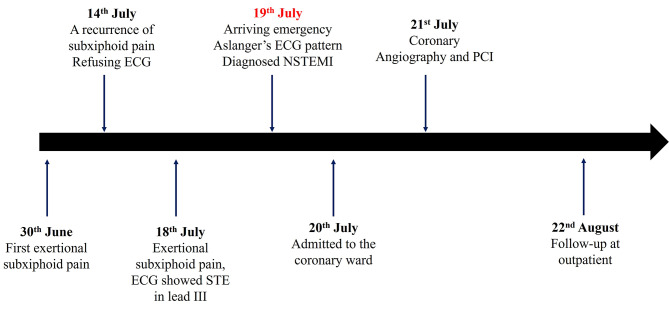
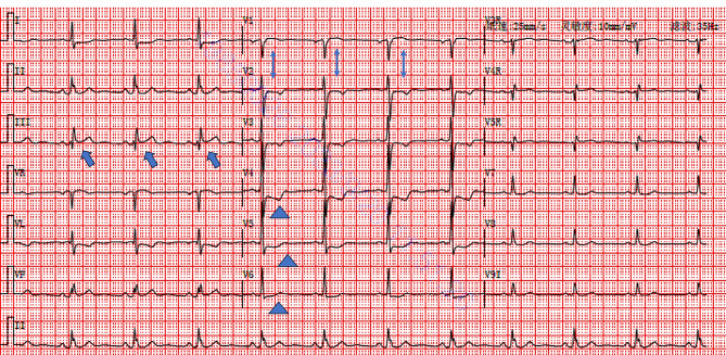
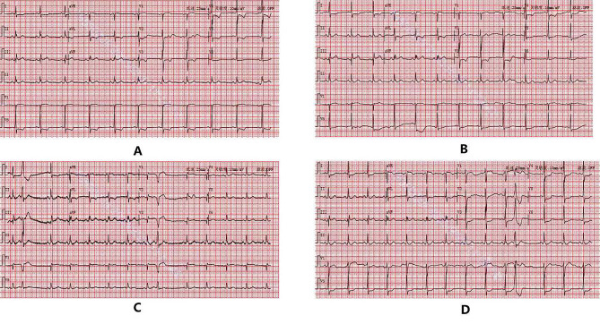
The patient was a 62-year-old male with chief complaint of intermittent exertional subxiphoid pain for 20 days from 30th June. One day after the last episode (19th July), the 18-lead electrocardiogram showed ST segment elevation of 0.05-0.1mV in lead III, ST segment depression in leads I, avL, and V2-V6, T wave inversion with positive terminal vector in lead V4-V5, and positive T wave in lead V6, which indicated Aslanger's pattern. With increased Troponin I (0.162ng/mL, 0-0.02), The patient was diagnosed as acute non-ST-segment elevation myocardial infarction (NSTEMI) and admitted to coronary ward on 20th July. The coronary angiography showed 95% stenosis in the distal left main coronary artery (LM) to the ostium of the left anterior descending artery (LAD), 90% stenosis in the proximal segment of the LAD, and 80% stenosis in the middle segment of the LAD, and TIMI blood flow was graded score 2. Three drug-eluting stents were implanted at the lesions. The patient's ECG returned close to normal one month after revascularization.

CONCLUSION

We presented an acute coronary syndrome case whose ECG showed with Aslanger's pattern (i.e., isolated ST-segment elevation in lead III, associated ST-segment depression in lead V4-V6 with positive T wave/terminal vector, and greater ST-segment elevation in lead V1 than in lead V2), and was confirmed severe stenosis of the LM and the proximal segment of the LAD via coronary angiography. In clinical practice, especially in the emergency, patients with ECG presenting Aslanger's pattern should be urgently evaluated with prompt treatment, and the timing of emergency coronary angiography and revascularization should be evaluated to avoid adverse outcomes caused by delayed treatment.

摘要

背景

心电图(ECG)中的 Aslanger 图形表明患者可能患有急性下壁心肌梗死(AMI),同时其他冠状动脉存在临界狭窄,需要评估血运重建的时机,将其视为 ST 段抬高型心肌梗死(STEMI)的等效风险。

病例介绍

患者,男性,62 岁,主因间断性胸骨下劳累性疼痛 20 天,于 6 月 30 日就诊。末次发作后 1 天(7 月 19 日),18 导联心电图显示 III 导联 ST 段抬高 0.05-0.1mV,I、avL 和 V2-V6 导联 ST 段压低,V4-V5 导联 T 波倒置,正向终末向量,V6 导联 T 波正向,提示 Aslanger 图形。肌钙蛋白 I 升高(0.162ng/ml,0-0.02),诊断为急性非 ST 段抬高型心肌梗死(NSTEMI),于 7 月 20 日收入心内科病房。冠状动脉造影显示左主干(LM)远段至左前降支(LAD)开口处狭窄 95%,LAD 近段狭窄 90%,中段狭窄 80%,TIMI 血流分级 2 级。于病变处植入 3 枚药物洗脱支架。血管重建术后 1 个月,患者心电图恢复接近正常。

结论

我们报告了 1 例急性冠状动脉综合征患者,其心电图呈 Aslanger 图形(即 III 导联孤立性 ST 段抬高,V4-V6 导联 ST 段压低伴正向 T 波/终末向量,V1 导联 ST 段抬高大于 V2 导联),经冠状动脉造影证实 LM 和 LAD 近段严重狭窄。在临床实践中,特别是在急诊情况下,出现心电图 Aslanger 图形的患者应紧急评估,及时治疗,并评估紧急冠状动脉造影和血运重建的时机,以避免因治疗延迟而导致不良后果。

https://cdn.ncbi.nlm.nih.gov/pmc/blobs/cc05/10763094/79746512ac3d/12872_2023_3678_Fig5_HTML.jpg
https://cdn.ncbi.nlm.nih.gov/pmc/blobs/cc05/10763094/1f3aed881260/12872_2023_3678_Fig1_HTML.jpg
https://cdn.ncbi.nlm.nih.gov/pmc/blobs/cc05/10763094/943bbee24920/12872_2023_3678_Fig2_HTML.jpg
https://cdn.ncbi.nlm.nih.gov/pmc/blobs/cc05/10763094/54cdc766c51e/12872_2023_3678_Fig3_HTML.jpg
https://cdn.ncbi.nlm.nih.gov/pmc/blobs/cc05/10763094/576f9b09d73b/12872_2023_3678_Fig4_HTML.jpg
https://cdn.ncbi.nlm.nih.gov/pmc/blobs/cc05/10763094/79746512ac3d/12872_2023_3678_Fig5_HTML.jpg

相似文献

1
A patient with acute myocardial infarction with electrocardiogram Aslanger's pattern.心电图表现为 Aslanger 样改变的急性心肌梗死患者。
BMC Cardiovasc Disord. 2024 Jan 2;24(1):3. doi: 10.1186/s12872-023-03678-x.
2
Clinical and angiographic characteristics of patients with combined anterior and inferior ST-segment elevation on the initial electrocardiogram during acute myocardial infarction.急性心肌梗死期间初始心电图表现为前壁和下壁ST段联合抬高患者的临床及血管造影特征
Am Heart J. 2003 Oct;146(4):653-61. doi: 10.1016/S0002-8703(03)00369-7.
3
Aslanger's pattern with acute lesion in the left coronary system: A case report.左冠状动脉系统急性病变的阿斯朗格氏型:一例报告。
J Electrocardiol. 2024 Nov-Dec;87:153807. doi: 10.1016/j.jelectrocard.2024.153807. Epub 2024 Sep 15.
4
The "criminal" artery of de Winter may be the left circumflex artery: A CARE-compliant case report.德温特“罪犯”动脉可能是左旋支动脉:一份符合CARE标准的病例报告。
Medicine (Baltimore). 2020 Jun 12;99(24):e20585. doi: 10.1097/MD.0000000000020585.
5
Value of the electrocardiogram in localizing the occlusion site in the left anterior descending coronary artery in acute anterior myocardial infarction.心电图在急性前壁心肌梗死中定位左前降支冠状动脉闭塞部位的价值。
J Am Coll Cardiol. 1999 Aug;34(2):389-95. doi: 10.1016/s0735-1097(99)00197-7.
6
Influence of the extent of coronary atherosclerotic disease on ST-segment changes induced by ST elevation myocardial infarction.冠状动脉粥样硬化病变程度对 ST 段抬高型心肌梗死诱导的 ST 段变化的影响。
Am J Cardiol. 2014 Mar 1;113(5):757-64. doi: 10.1016/j.amjcard.2013.11.034. Epub 2013 Dec 12.
7
Acute diagonal-induced ST-elevation myocardial infarction and electrocardiogram-guidance in the era of primary coronary intervention: New insights into an old tool.急性斜角诱导的 ST 段抬高型心肌梗死和直接经皮冠状动脉介入治疗时代的心电图指导:老工具的新见解。
Eur Heart J Acute Cardiovasc Care. 2020 Dec;9(8):827-835. doi: 10.1177/2048872619828291. Epub 2019 Feb 1.
8
A new 4-variable formula to differentiate normal variant ST segment elevation in V2-V4 (early repolarization) from subtle left anterior descending coronary occlusion - Adding QRS amplitude of V2 improves the model.一种用于区分V2-V4导联正常变异ST段抬高(早期复极)与轻微左前降支冠状动脉闭塞的新四变量公式——增加V2导联的QRS波振幅可改善该模型。
J Electrocardiol. 2017 Sep-Oct;50(5):561-569. doi: 10.1016/j.jelectrocard.2017.04.005. Epub 2017 Apr 19.
9
Value of T-wave direction with lead III ST-segment depression in acute anterior wall myocardial infarction: electrocardiographic prediction of a "wrapped" left anterior descending artery.急性前壁心肌梗死时Ⅲ导联ST段压低伴T波方向的价值:“包绕型”左前降支的心电图预测
Clin Cardiol. 1998 Aug;21(8):562-6. doi: 10.1002/clc.4960210806.
10
A Case of ST-Elevation Myocardial Infarction With Right Bundle Branch Block, an Ominous Sign of Critical Coronary Occlusion.一例伴有右束支传导阻滞的ST段抬高型心肌梗死,冠状动脉严重闭塞的不祥征兆。
Cureus. 2022 Jan 13;14(1):e21216. doi: 10.7759/cureus.21216. eCollection 2022 Jan.

本文引用的文献

1
A proposal for better visualization of Aslanger pattern.一种改进 Aslanger 模式可视化效果的建议。
J Electrocardiol. 2023 Nov-Dec;81:41-43. doi: 10.1016/j.jelectrocard.2023.07.009. Epub 2023 Jul 24.
2
Clinical Features of the Aslanger Pattern to Compensate for the Limitation of ST-Elevation Myocardial Infarction (STEMI) Criteria.用于弥补ST段抬高型心肌梗死(STEMI)标准局限性的阿斯朗格模式的临床特征。
Cureus. 2023 Jan 1;15(1):e33227. doi: 10.7759/cureus.33227. eCollection 2023 Jan.
3
Acute inferior occlusion myocardial infarction with a solitary ST-elevation in lead III: A case report.
急性下壁伴 III 导联孤立性 ST 段抬高型心肌梗死:1 例报告。
J Electrocardiol. 2022 May-Jun;72:35-38. doi: 10.1016/j.jelectrocard.2022.02.010. Epub 2022 Feb 28.
4
The significance of the morphology-voltage-P-wave duration (MVP) ECG score for prediction of in-hospital and long-term atrial fibrillation in ischemic stroke.形态学-电压-P波时限(MVP)心电图评分对预测缺血性卒中患者住院期间及长期房颤的意义
J Electrocardiol. 2021 Nov-Dec;69:44-50. doi: 10.1016/j.jelectrocard.2021.09.006. Epub 2021 Sep 14.
5
Recognizing electrocardiographically subtle occlusion myocardial infarction and differentiating it from mimics: Ten steps to or away from cath lab.识别心电图细微闭塞性心肌梗死并将其与模拟病例区分开来:走向或远离心导管实验室的十步。
Turk Kardiyol Dern Ars. 2021 Sep;49(6):488-500. doi: 10.5543/tkda.2021.21026.
6
A simple formula to predict echocardiographic diastolic dysfunction-electrocardiographic diastolic index.一种预测超声心动图舒张功能障碍的简单公式-心电图舒张指数。
Herz. 2021 Sep;46(Suppl 2):159-165. doi: 10.1007/s00059-020-04972-6. Epub 2020 Aug 10.
7
In-Hospital Prognostic Value of Electrocardiographic Parameters Other Than ST-Segment Changes in Acute Myocardial Infarction: Literature Review and Future Perspectives.急性心肌梗死心电图 ST 段改变以外的电生理参数的院内预后价值:文献回顾与未来展望。
Heart Lung Circ. 2020 Nov;29(11):1603-1612. doi: 10.1016/j.hlc.2020.04.011. Epub 2020 Jun 11.
8
A new electrocardiographic pattern indicating inferior myocardial infarction.一种提示下壁心肌梗死的新心电图模式。
J Electrocardiol. 2020 Jul-Aug;61:41-46. doi: 10.1016/j.jelectrocard.2020.04.008. Epub 2020 Apr 13.