School of Biomedical Sciences, Faculty of Biological Sciences, University of Leeds, Leeds LS2 9JT, UK.
School of Biosciences, Faculty of Science, The University of Sheffield, Sheffield S10 2TN, UK.
Cells. 2023 Dec 23;13(1):38. doi: 10.3390/cells13010038.
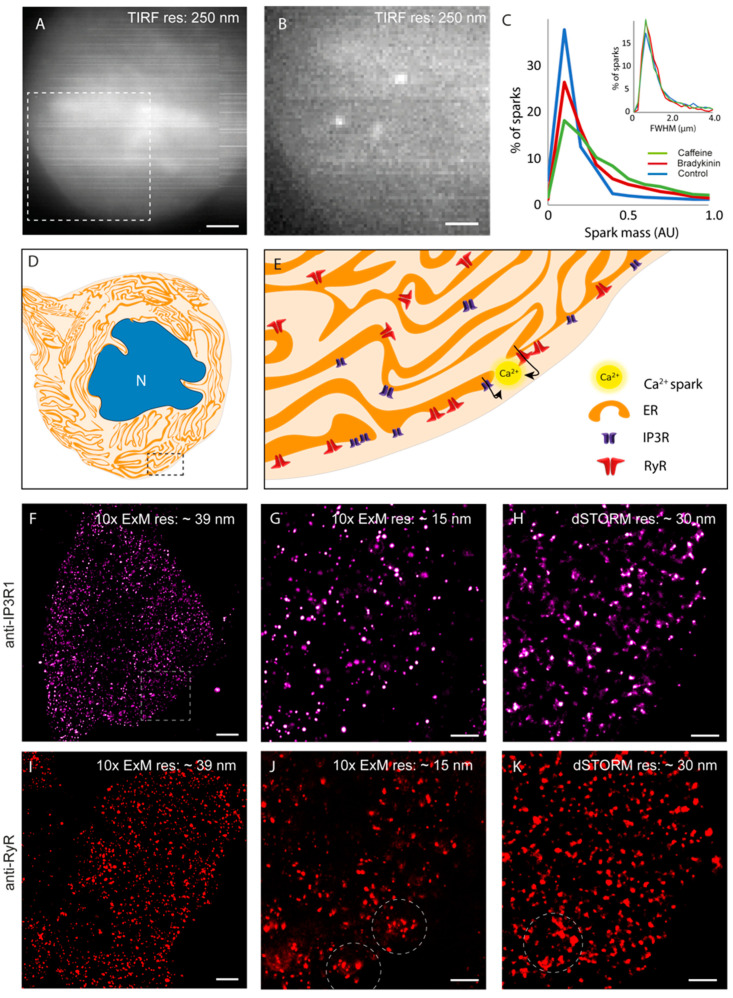
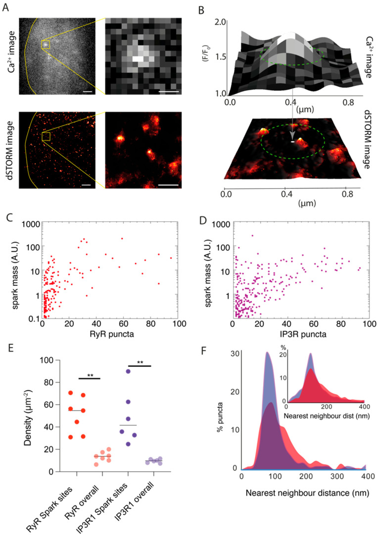
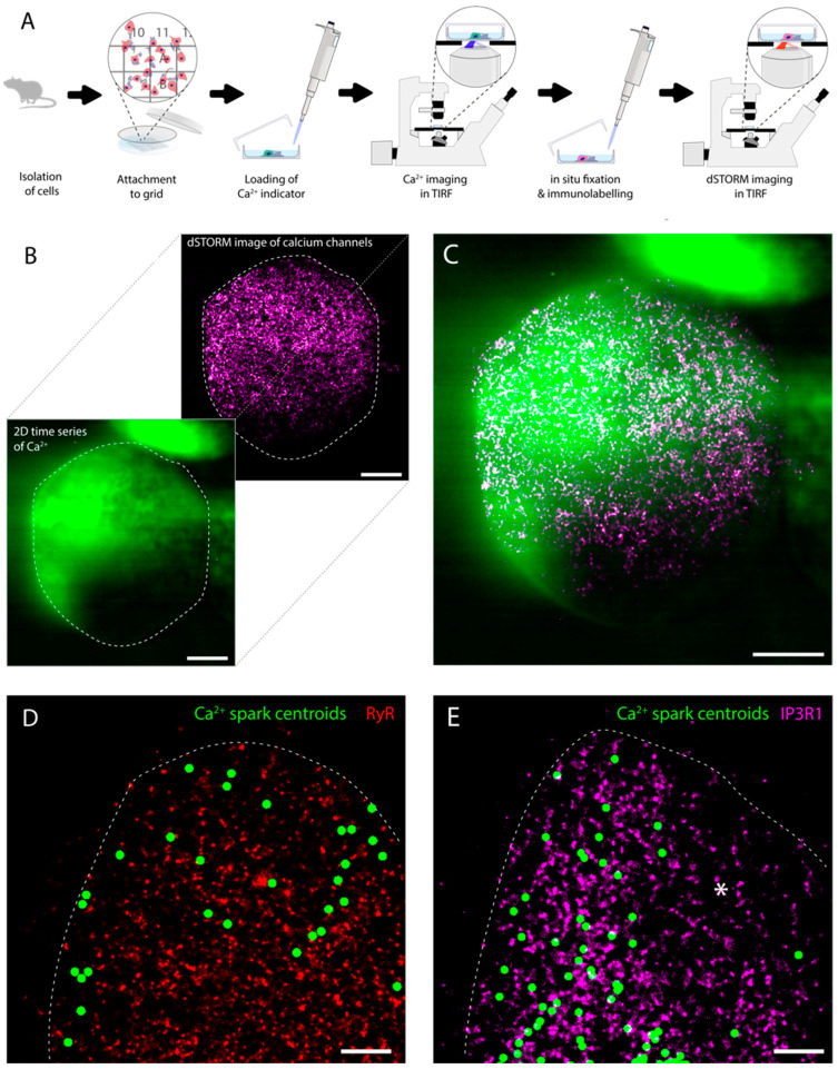
Coordinated events of calcium (Ca) released from the endoplasmic reticulum (ER) are key second messengers in excitable cells. In pain-sensing dorsal root ganglion (DRG) neurons, these events can be observed as Ca sparks, produced by a combination of ryanodine receptors (RyR) and inositol 1,4,5-triphosphate receptors (IP3R1). These microscopic signals offer the neuronal cells with a possible means of modulating the subplasmalemmal Ca handling, initiating vesicular exocytosis. With super-resolution dSTORM and expansion microscopies, we visualised the nanoscale distributions of both RyR and IP3R1 that featured loosely organised clusters in the subplasmalemmal regions of cultured rat DRG somata. We adapted a novel correlative microscopy protocol to examine the nanoscale patterns of RyR and IP3R1 in the locality of each Ca spark. We found that most subplasmalemmal sparks correlated with relatively small groups of RyR whilst larger sparks were often associated with larger groups of IP3R1. These data also showed spontaneous Ca sparks in <30% of the subplasmalemmal cell area but consisted of both these channel species at a 3.8-5 times higher density than in nonactive regions of the cell. Taken together, these observations reveal distinct patterns and length scales of RyR and IP3R1 co-clustering at contact sites between the ER and the surface plasmalemma that encode the positions and the quantity of Ca released at each Ca spark.
钙(Ca)从内质网(ER)中释放出来的协调事件是兴奋性细胞中的关键第二信使。在痛觉感觉背根神经节(DRG)神经元中,这些事件可以观察到 Ca 火花,这是由兰尼碱受体(RyR)和肌醇 1,4,5-三磷酸受体(IP3R1)的组合产生的。这些微观信号为神经元细胞提供了一种调节质膜下 Ca 处理的可能方式,从而引发囊泡胞吐。通过超分辨率 dSTORM 和扩展显微镜,我们可视化了 RyR 和 IP3R1 的纳米级分布,这些分布在培养的大鼠 DRG 体神经元的质膜下区域具有松散组织的簇。我们采用了一种新的相关显微镜方案来检查 RyR 和 IP3R1 在每个 Ca 火花附近的纳米级模式。我们发现,大多数质膜下火花与相对较小的 RyR 群相关,而较大的火花通常与较大的 IP3R1 群相关。这些数据还表明,质膜下细胞区域 <30%的自发 Ca 火花包含这两种通道,但它们的密度比细胞非活动区高 3.8-5 倍。综上所述,这些观察结果揭示了 RyR 和 IP3R1 在 ER 和质膜表面之间的接触点处的明显的聚类模式和长度尺度,这些模式和长度尺度编码了每个 Ca 火花中 Ca 释放的位置和数量。