• 文献检索
  • 文档翻译
  • 深度研究
  • 学术资讯
  • Suppr Zotero 插件Zotero 插件
  • 邀请有礼
  • 套餐&价格
  • 历史记录
应用&插件
Suppr Zotero 插件Zotero 插件浏览器插件Mac 客户端Windows 客户端微信小程序
定价
高级版会员购买积分包购买API积分包
服务
文献检索文档翻译深度研究API 文档MCP 服务
关于我们
关于 Suppr公司介绍联系我们用户协议隐私条款
关注我们

Suppr 超能文献

核心技术专利:CN118964589B侵权必究
粤ICP备2023148730 号-1Suppr @ 2026

文献检索

告别复杂PubMed语法,用中文像聊天一样搜索,搜遍4000万医学文献。AI智能推荐,让科研检索更轻松。

立即免费搜索

文件翻译

保留排版,准确专业,支持PDF/Word/PPT等文件格式,支持 12+语言互译。

免费翻译文档

深度研究

AI帮你快速写综述,25分钟生成高质量综述,智能提取关键信息,辅助科研写作。

立即免费体验

质膜起泡受波形蛋白中间丝亚细胞分布控制。

Plasma Membrane Blebbing Is Controlled by Subcellular Distribution of Vimentin Intermediate Filaments.

机构信息

N.N. Blokhin National Medical Research Center of Oncology, 24 Kashirskoe Shosse, Moscow 115478, Russia.

Dynamics of Immune Responses Team, INSERM-U1223 Institut Pasteur, 25-28 Rue du Dr Roux, 75015 Paris, France.

出版信息

Cells. 2024 Jan 4;13(1):105. doi: 10.3390/cells13010105.

DOI:10.3390/cells13010105
PMID:38201309
原文链接:https://pmc.ncbi.nlm.nih.gov/articles/PMC10778383/
Abstract

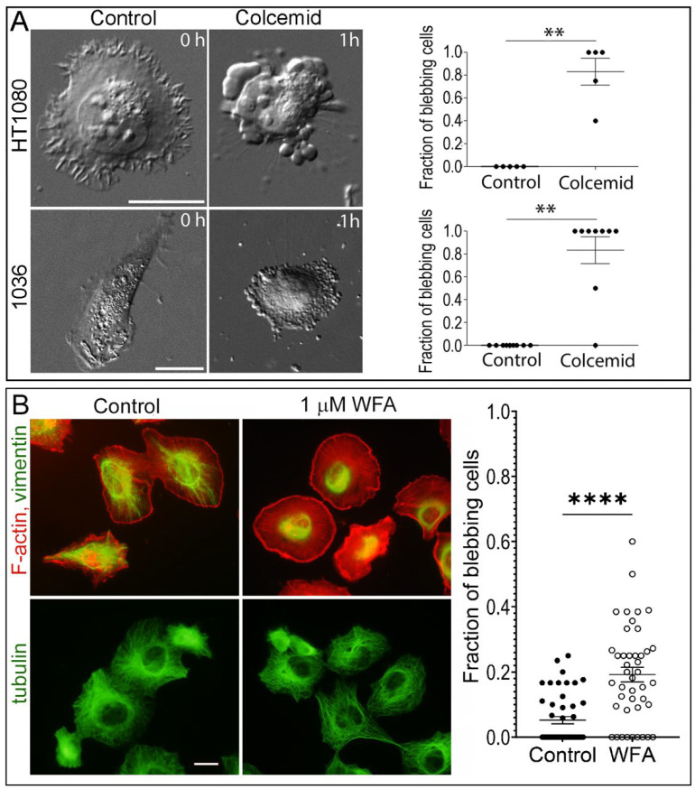
The formation of specific cellular protrusions, plasma membrane blebs, underlies the amoeboid mode of cell motility, which is characteristic for free-living amoebae and leukocytes, and can also be adopted by stem and tumor cells to bypass unfavorable migration conditions and thus facilitate their long-distance migration. Not all cells are equally prone to bleb formation. We have previously shown that membrane blebbing can be experimentally induced in a subset of HT1080 fibrosarcoma cells, whereas other cells in the same culture under the same conditions retain non-blebbing mesenchymal morphology. Here we show that this heterogeneity is associated with the distribution of vimentin intermediate filaments (VIFs). Using different approaches to alter the VIF organization, we show that blebbing activity is biased toward cell edges lacking abundant VIFs, whereas the VIF-rich regions of the cell periphery exhibit low blebbing activity. This pattern is observed both in interphase fibroblasts, with and without experimentally induced blebbing, and during mitosis-associated blebbing. Moreover, the downregulation of vimentin expression or displacement of VIFs away from the cell periphery promotes blebbing even in cells resistant to bleb-inducing treatments. Thus, we reveal a new important function of VIFs in cell physiology that involves the regulation of non-apoptotic blebbing essential for amoeboid cell migration and mitosis.

摘要

特定细胞突起的形成,即质膜泡,是变形虫样运动细胞的运动方式的基础,这种运动方式的特点是自由生活的变形虫和白细胞,也可以被干细胞和肿瘤细胞采用,以绕过不利的迁移条件,从而促进其远距离迁移。并非所有细胞都同样容易形成质膜泡。我们之前已经表明,可以在 HT1080 纤维肉瘤细胞的亚群中实验性诱导质膜泡形成,而在相同条件下同一培养物中的其他细胞则保持非泡状的间充质形态。在这里,我们表明这种异质性与波形蛋白中间丝(VIFs)的分布有关。使用改变 VIF 组织的不同方法,我们表明质膜泡活性偏向于缺乏丰富 VIF 的细胞边缘,而细胞边缘富含 VIF 的区域则表现出低质膜泡活性。这种模式在有丝分裂前期的成纤维细胞中均有观察到,无论是在实验诱导质膜泡形成的细胞中,还是在与有丝分裂相关的质膜泡形成的细胞中。此外,下调波形蛋白的表达或使 VIF 从细胞边缘移位,甚至可以促进那些对诱导质膜泡形成的处理有抗性的细胞发生质膜泡形成。因此,我们揭示了 VIF 在细胞生理学中的一个新的重要功能,它涉及到非凋亡性质膜泡的调节,这对于变形虫样细胞的迁移和有丝分裂是必不可少的。

https://cdn.ncbi.nlm.nih.gov/pmc/blobs/a82d/10778383/826c9359f1e5/cells-13-00105-g006.jpg
https://cdn.ncbi.nlm.nih.gov/pmc/blobs/a82d/10778383/6f022dd4fd4a/cells-13-00105-g001.jpg
https://cdn.ncbi.nlm.nih.gov/pmc/blobs/a82d/10778383/9f9522fdf649/cells-13-00105-g002.jpg
https://cdn.ncbi.nlm.nih.gov/pmc/blobs/a82d/10778383/ad9dbf569e2d/cells-13-00105-g003.jpg
https://cdn.ncbi.nlm.nih.gov/pmc/blobs/a82d/10778383/49a1344c8c13/cells-13-00105-g004.jpg
https://cdn.ncbi.nlm.nih.gov/pmc/blobs/a82d/10778383/645340466c6d/cells-13-00105-g005.jpg
https://cdn.ncbi.nlm.nih.gov/pmc/blobs/a82d/10778383/826c9359f1e5/cells-13-00105-g006.jpg

相似文献

1
Plasma Membrane Blebbing Is Controlled by Subcellular Distribution of Vimentin Intermediate Filaments.质膜起泡受波形蛋白中间丝亚细胞分布控制。
Cells. 2024 Jan 4;13(1):105. doi: 10.3390/cells13010105.
2
Impairment of Assembly of the Vimentin Intermediate Filaments Leads to Suppression of Formation and Maturation of Focal Contacts and Alteration of the Type of Cellular Protrusions.中间丝角蛋白组装的损伤导致黏着斑的形成和成熟受到抑制,并改变细胞突起的类型。
Biochemistry (Mosc). 2024 Jan;89(1):184-195. doi: 10.1134/S0006297924010127.
3
Transition from mesenchymal to bleb-based motility is predominantly exhibited by CD133-positive subpopulation of fibrosarcoma cells.间充质样到小泡样运动的转变主要由纤维肉瘤细胞中 CD133 阳性亚群表现出来。
Biol Cell. 2019 Oct;111(10):245-261. doi: 10.1111/boc.201800078. Epub 2019 Aug 22.
4
Vimentin intermediate filaments and filamentous actin form unexpected interpenetrating networks that redefine the cell cortex.波形蛋白中间纤维和丝状肌动蛋白形成意想不到的贯穿网络,重新定义了细胞皮质。
Proc Natl Acad Sci U S A. 2022 Mar 8;119(10):e2115217119. doi: 10.1073/pnas.2115217119. Epub 2022 Mar 2.
5
A flexible network of vimentin intermediate filaments promotes migration of amoeboid cancer cells through confined environments.波形蛋白中间丝构成的柔性网络促进了阿米巴样癌细胞在受限环境中的迁移。
J Biol Chem. 2020 May 8;295(19):6700-6709. doi: 10.1074/jbc.RA119.011537. Epub 2020 Mar 31.
6
Vimentin Intermediate Filaments Template Microtubule Networks to Enhance Persistence in Cell Polarity and Directed Migration.波形蛋白中间丝模板微管网络增强细胞极性和定向迁移中的持久性。
Cell Syst. 2016 Sep 28;3(3):252-263.e8. doi: 10.1016/j.cels.2016.08.007. Epub 2016 Sep 22.
7
Met-induced membrane blebbing leads to amoeboid cell motility and invasion.金属诱导的细胞膜起泡导致变形虫样细胞的运动和侵袭。
Oncogene. 2014 Apr 3;33(14):1788-98. doi: 10.1038/onc.2013.138. Epub 2013 May 13.
8
Src and SHP2 coordinately regulate the dynamics and organization of vimentin filaments during cell migration.Src 和 SHP2 协同调节细胞迁移过程中中间丝蛋白(vimentin)纤维的动态和组织。
Oncogene. 2019 May;38(21):4075-4094. doi: 10.1038/s41388-019-0705-x. Epub 2019 Jan 29.
9
Atypical PKCs activate Vimentin to facilitate prostate cancer cell motility and invasion.非典型蛋白激酶 C 激活波形蛋白以促进前列腺癌细胞迁移和侵袭。
Cell Adh Migr. 2021 Dec;15(1):37-57. doi: 10.1080/19336918.2021.1882782.
10
Vimentin filaments drive migratory persistence in polyploidal cancer cells.中间丝蛋白丝驱动多倍体癌细胞的迁移持续。
Proc Natl Acad Sci U S A. 2020 Oct 27;117(43):26756-26765. doi: 10.1073/pnas.2011912117. Epub 2020 Oct 12.

引用本文的文献

1
Genetic Animal Models of Idiopathic Generalized Epilepsies: What Can We Learn from Them?特发性全身性癫痫的遗传动物模型:我们能从中学到什么?
Biomedicines. 2025 May 26;13(6):1301. doi: 10.3390/biomedicines13061301.
2
Potential and value of rescuing dying neurons.拯救濒死神经元的潜力与价值
Neural Regen Res. 2026 Mar 1;21(3):1013-1022. doi: 10.4103/NRR.NRR-D-24-01134. Epub 2025 Apr 29.
3
Three-dimensional cell culture conditions promoted the Mesenchymal-Amoeboid Transition in the Triple-Negative Breast Cancer cell line MDA-MB-231.

本文引用的文献

1
Vimentin intermediate filaments and filamentous actin form unexpected interpenetrating networks that redefine the cell cortex.波形蛋白中间纤维和丝状肌动蛋白形成意想不到的贯穿网络,重新定义了细胞皮质。
Proc Natl Acad Sci U S A. 2022 Mar 8;119(10):e2115217119. doi: 10.1073/pnas.2115217119. Epub 2022 Mar 2.
2
A flexible network of vimentin intermediate filaments promotes migration of amoeboid cancer cells through confined environments.波形蛋白中间丝构成的柔性网络促进了阿米巴样癌细胞在受限环境中的迁移。
J Biol Chem. 2020 May 8;295(19):6700-6709. doi: 10.1074/jbc.RA119.011537. Epub 2020 Mar 31.
3
F-Actin Interactome Reveals Vimentin as a Key Regulator of Actin Organization and Cell Mechanics in Mitosis.
三维细胞培养条件促进了三阴性乳腺癌细胞系MDA-MB-231中的间充质-阿米巴样转变。
Front Cell Dev Biol. 2024 Aug 2;12:1435708. doi: 10.3389/fcell.2024.1435708. eCollection 2024.
F 肌动蛋白相互作用组揭示波形蛋白作为有丝分裂中肌动蛋白组织和细胞力学的关键调节剂。
Dev Cell. 2020 Jan 27;52(2):210-222.e7. doi: 10.1016/j.devcel.2019.12.011. Epub 2020 Jan 9.
4
Loss of Vimentin Enhances Cell Motility through Small Confining Spaces.波形蛋白缺失通过狭小的封闭空间增强细胞迁移能力。
Small. 2019 Dec;15(50):e1903180. doi: 10.1002/smll.201903180. Epub 2019 Nov 13.
5
Partial loss of actin nucleator actin-related protein 2/3 activity triggers blebbing in primary T lymphocytes.肌动蛋白成核因子肌动蛋白相关蛋白 2/3 活性部分缺失引发原代 T 淋巴细胞起泡。
Immunol Cell Biol. 2020 Feb;98(2):93-113. doi: 10.1111/imcb.12304. Epub 2019 Dec 23.
6
Vimentin filaments interact with the actin cortex in mitosis allowing normal cell division.波形蛋白丝在有丝分裂中与肌动蛋白皮质相互作用,从而允许正常的细胞分裂。
Nat Commun. 2019 Sep 13;10(1):4200. doi: 10.1038/s41467-019-12029-4.
7
Transition from mesenchymal to bleb-based motility is predominantly exhibited by CD133-positive subpopulation of fibrosarcoma cells.间充质样到小泡样运动的转变主要由纤维肉瘤细胞中 CD133 阳性亚群表现出来。
Biol Cell. 2019 Oct;111(10):245-261. doi: 10.1111/boc.201800078. Epub 2019 Aug 22.
8
A mechano-signalling network linking microtubules, myosin IIA filaments and integrin-based adhesions.一个机械信号网络,将微管、肌球蛋白 IIA 丝和整合素基黏附连接起来。
Nat Mater. 2019 Jun;18(6):638-649. doi: 10.1038/s41563-019-0371-y. Epub 2019 May 21.
9
Cancer Cells Invade Confined Microchannels via a Self-Directed Mesenchymal-to-Amoeboid Transition.癌细胞通过自我导向的间质-阿米巴样转变侵袭受限的微通道。
Nano Lett. 2019 Apr 10;19(4):2280-2290. doi: 10.1021/acs.nanolett.8b04720. Epub 2019 Mar 8.
10
Time-resolved ultrastructure of the cortical actin cytoskeleton in dynamic membrane blebs.动态质膜泡中皮质肌动蛋白细胞骨架的时间分辨超微结构。
J Cell Biol. 2019 Feb 4;218(2):445-454. doi: 10.1083/jcb.201806075. Epub 2018 Dec 12.