Division of Neuroimmunology, Joint Research Center for Human Retrovirus Infection, Kagoshima University, 8-35-1 Sakuragaoka, Kagoshima 890-8544, Japan.
Faculty of Bioscience, Nagahama Institute of Bio-Science and Technology, 1266 Tamura, Nagahama 526-0829, Japan.
Int J Mol Sci. 2023 Dec 27;25(1):383. doi: 10.3390/ijms25010383.
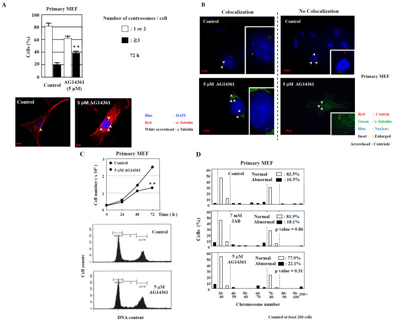
The centrosome is involved in cytoplasmic microtubule organization during interphase and in mitotic spindle assembly during cell division. Centrosome amplification (abnormal proliferation of centrosome number) has been observed in several types of cancer and in precancerous conditions. Therefore, it is important to elucidate the mechanism of centrosome amplification in order to understand the early stage of carcinogenesis. Primary cells could be used to better understand the early stage of carcinogenesis rather than immortalized cells, which tend to have various genetic and epigenetic changes. Previously, we demonstrated that a poly(ADP-ribose) polymerase (PARP) inhibitor, 3-aminobenzamide (3AB), which is known to be nontoxic and nonmutagenic, could induce centrosome amplification and chromosomal aneuploidy in CHO-K1 cells. In this study, we compared primary mouse embryonic fibroblasts (MEF) and immortalized MEF using 3AB. Although centrosome amplification was induced with 3AB treatment in immortalized MEF, a more potent PARP inhibitor, AG14361, was required for primary MEF. However, after centrosome amplification, neither 3AB in immortalized MEF nor AG14361 in primary MEF caused chromosomal aneuploidy, suggesting that further genetic and/or epigenetic change(s) are required to exhibit aneuploidy. The DNA-damaging agents doxorubicin and γ-irradiation can cause cancer and centrosome amplification in experimental animals. Although doxorubicin and γ-irradiation induced centrosome amplification and led to decreased p27Kip protein levels in immortalized MEF and primary MEF, the phosphorylation ratio of nucleophosmin (Thr199) increased in immortalized MEF, whereas it decreased in primary MEF. These results suggest that there exists a yet unidentified pathway, different from the nucleophosmin phosphorylation pathway, which can cause centrosome amplification in primary MEF.
中心体参与间期细胞质微管组织和细胞分裂时有丝分裂纺锤体的组装。在几种类型的癌症和癌前状态中观察到中心体扩增(中心体数量异常增殖)。因此,阐明中心体扩增的机制对于了解癌变的早期阶段非常重要。与容易发生各种遗传和表观遗传改变的永生化细胞相比,原代细胞可以更好地理解癌变的早期阶段。以前,我们证明聚(ADP-核糖)聚合酶(PARP)抑制剂 3-氨基苯甲酰胺(3AB)可诱导 CHO-K1 细胞的中心体扩增和染色体非整倍性。在这项研究中,我们使用 3AB 比较了原代小鼠胚胎成纤维细胞(MEF)和永生化 MEF。虽然 3AB 处理诱导了永生化 MEF 的中心体扩增,但需要更有效的 PARP 抑制剂 AG14361 才能诱导原代 MEF 的中心体扩增。然而,在中心体扩增后,无论是在永生化 MEF 中使用 3AB 还是在原代 MEF 中使用 AG14361,都不会导致染色体非整倍性,这表明需要进一步的遗传和/或表观遗传改变才能表现出非整倍性。阿霉素和γ射线等 DNA 损伤剂可在实验动物中引起癌症和中心体扩增。尽管阿霉素和γ射线诱导了中心体扩增,并导致永生化 MEF 和原代 MEF 中 p27Kip 蛋白水平降低,但核仁磷酸蛋白(Thr199)的磷酸化比例在永生化 MEF 中增加,而在原代 MEF 中则降低。这些结果表明,存在一种尚未确定的途径,不同于核仁磷酸蛋白磷酸化途径,可导致原代 MEF 中的中心体扩增。