Ahmed Nasar, Khalil Zakia, Farooq Zahid, Shahida Shabnam, Ahmad Pervaiz, Qadir Karwan Wasman, Khan Rajwali, Zafar Qayyum
Department of Physics, King Abdullah Campus, University of Azad Jammu and Kashmir, Muzaffarabad 13100, Pakistan.
Department of Physics, Mirpur University of Science and Technology, Muzaffarabad, Azad Jammu and Kashmir 10250, Pakistan.
ACS Omega. 2023 Dec 19;9(1):137-145. doi: 10.1021/acsomega.3c01727. eCollection 2024 Jan 9.
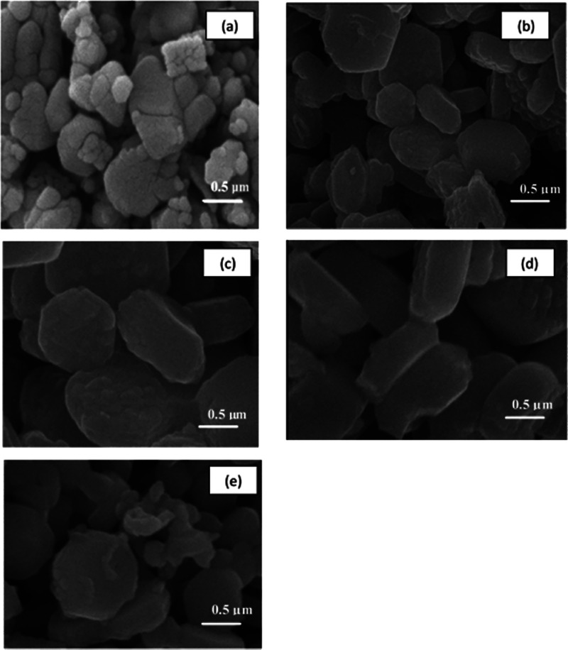
Pure and Ni-Fe-codoped ZnNiFeO ( = 0.01, 0.02, 0.03, and 0.04) nanoparticles were effectively synthesized using a sol-gel autocombustion procedure. The structural, optical, morphological, and magnetic properties were determined by using X-ray diffraction (XRD), ultraviolet-visible (UV-vis), scanning electron microscopy, and vibrating sample magnetometer techniques. The XRD confirmed the purity of the hexagonal wurtzite crystal structure. XRD analysis further indicated that Fe and Ni successfully substituted the lattice site of Zn and generated a single-phase ZnNiFeO magnetic oxide. In addition, a significant morphological change was observed with an increase in the dopant concentration by using high-resolution scanning electron microscopy. The UV-vis spectroscopy analysis indicated the redshift in the optical band gap with increasing dopant concentration signifying a progressive decrease in the optical band gap. The vibrating sample magnetometer analysis revealed that the doped samples exhibited ferromagnetic properties at room temperature with an increase in the dopant concentration. Dopant concentration was confirmed by using energy-dispersive X-ray spectroscopy. The current results provide a vital method to improve the magnetic properties of ZnO nanoparticles, which may get significant attention from researchers in the field of magnetic semiconductors.
采用溶胶-凝胶自燃烧法有效合成了纯的以及镍铁共掺杂的ZnNiFeO(= 0.01、0.02、0.03和0.04)纳米颗粒。利用X射线衍射(XRD)、紫外可见(UV-vis)、扫描电子显微镜和振动样品磁强计技术测定了其结构、光学、形态和磁性。XRD证实了六方纤锌矿晶体结构的纯度。XRD分析进一步表明,铁和镍成功取代了锌的晶格位置,生成了单相ZnNiFeO磁性氧化物。此外,通过高分辨率扫描电子显微镜观察到,随着掺杂剂浓度的增加,形态发生了显著变化。UV-vis光谱分析表明,随着掺杂剂浓度的增加,光学带隙出现红移,这意味着光学带隙逐渐减小。振动样品磁强计分析表明,掺杂样品在室温下表现出铁磁性能,且随着掺杂剂浓度的增加而增强。通过能量色散X射线光谱法确定了掺杂剂浓度。目前的结果提供了一种改善ZnO纳米颗粒磁性的重要方法,这可能会引起磁半导体领域研究人员的极大关注。