Gupta Anubhavv, Singh Jaspal, Mishra Atul, Singla Sanjeev K, Singh Ravinder Pal, Nar Amandeep Singh, Bawa Ashvind
Department of Surgery, Dayanand Medical College and Hospital, Ludhiana, Punjab, India.
J Minim Access Surg. 2024 Jan 1;20(1):89-95. doi: 10.4103/jmas.jmas_228_22. Epub 2023 Oct 18.
The most dreaded complication during laparoscopic cholecystectomy still remains to be injury to the common bile duct. The primary cause for bile duct injury during LC is misinterpretation of the biliary anatomy. Intra-operative cholangiography was introduced as a means of reducing the chances of biliary injury, done using Fluoroscopic imaging or Near-infrared fluorescence imaging method. NIRF is one of the most popular imaging methods in biomedical sciences. Indocyanine Green is sterile and water soluble which completely binds to albumin and is excreted in bile.
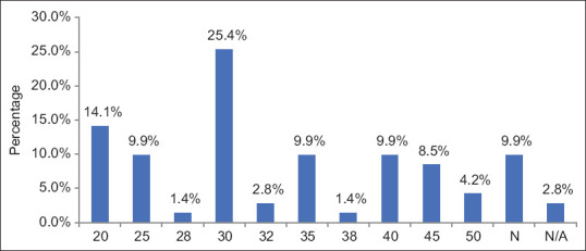
This prospective study was conducted among 70 patients between July 2020 and December 2021. Subjects were administered 5mg of ICG dye pre-operatively and procedure performed using Karl Storz HD image S1 system with a D-light P light source for NIRF imaging.
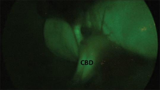
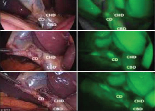
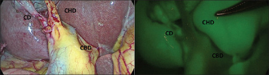
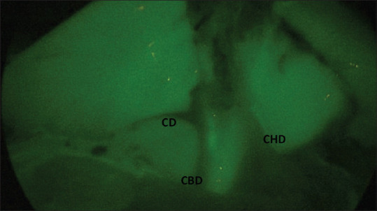
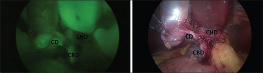
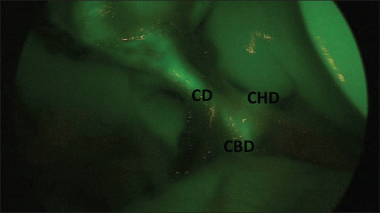
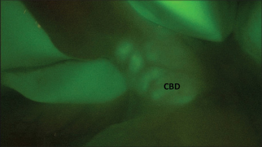
The average duration of surgery was 58.10 minutes. After calot's dissection, the CBD was visualized in 88.71 % patients, with a mean time to visualization at 26.33 minutes. The cystic duct was visualized in 87.3% cases with a mean time of visualization of 32.10 minutes. The hepatic duct was visualized in 28.57% and the hepatic duct-CBD confluence was visualized in 34.28% patients.
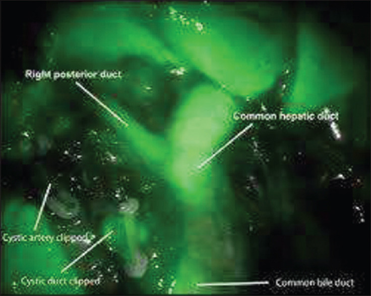
Near infrared imaging based intra-operative cholangiography, using Indocyanine Green dye, during Lap. Cholecystectomy is an easy, useful and inexpensive method of visualizing the biliary ductal anatomy.
腹腔镜胆囊切除术中最可怕的并发症仍然是胆总管损伤。腹腔镜胆囊切除术期间胆管损伤的主要原因是对胆管解剖结构的错误解读。术中胆管造影作为一种减少胆管损伤几率的方法被引入,可通过荧光透视成像或近红外荧光成像方法进行。近红外荧光成像(NIRF)是生物医学科学中最常用的成像方法之一。吲哚菁绿无菌且水溶性好,它完全与白蛋白结合并通过胆汁排泄。
这项前瞻性研究于2020年7月至2021年12月期间对70例患者进行。术前给受试者注射5mg吲哚菁绿染料,并使用卡尔·史托斯高清图像S1系统及D-light P光源进行近红外荧光成像手术。
平均手术时长为58.10分钟。在胆囊三角解剖后,88.71%的患者可见胆总管,平均可视化时间为26.33分钟。87.3%的病例可见胆囊管,平均可视化时间为32.10分钟。28.57%的患者可见肝管,34.28%的患者可见肝管-胆总管汇合处。
在腹腔镜胆囊切除术中,使用吲哚菁绿染料进行基于近红外成像的术中胆管造影是一种可视化胆管解剖结构的简单、实用且经济的方法。