Chemtob Erica V, Lin Dora H, Lee Esther, Heinz Eric R
School of Medicine and Health Sciences, The George Washington University, Washington, DC, United States.
Department of Anesthesiology and Critical Care Medicine, George Washington School of Medicine and Health Sciences, Washington, DC, United States.
J Anaesthesiol Clin Pharmacol. 2023 Oct-Dec;39(4):583-586. doi: 10.4103/joacp.joacp_113_22. Epub 2022 Sep 2.
Our study aimed to use submandibular ultrasound to measure upper airway parameters before and after induction dose of propofol in order to further understand upper airway changes that occur during induction of anesthesia. Measuring the changes that occur in airway anatomy due to the hypotonic effects of induction agents will allow for a deeper understanding of airway management.
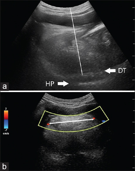
We enrolled 39 patients between November 2021 and January 2022. Submandibular ultrasound was used to measure tongue thickness, geniohyoid muscle thickness, the distance between the lingual arteries (DLA), lateral pharyngeal wall thickness, and hyomental distance before and after administration of induction doses of the commonly used, sedative-hypnotic agent, propofol.
The mean DLA increased significantly after propofol administration, from 3.62 ± 0.63 cm to 3.79 ± 0.56 cm ( < 0.001). The mean tongue thickness was 4.89 ± 0.51 cm and decreased significantly to a mean of 4.62 ± 0.50 cm after propofol administration ( < 0.001). The change in DLA measurements after propofol administration decreased significantly as STOP-BANG score increased (r = -0.344, = 0.037). However, DLA measurements when patients were awake increased significantly with an increase in the STOP-BANG score (r = 0.351, = 0.031).
These findings suggest that propofol widens and flattens the tongue, which are changes that may contribute to difficult airway management. Given the quick and non-invasive nature of ultrasound, further studies should evaluate the role of submandibular ultrasound for understanding the upper airway and airway management in various populations.
我们的研究旨在使用下颌下超声测量异丙酚诱导剂量给药前后的上气道参数,以进一步了解麻醉诱导期间发生的上气道变化。测量诱导药物的低张作用引起的气道解剖结构变化将有助于更深入地了解气道管理。
我们在2021年11月至2022年1月期间招募了39名患者。使用下颌下超声测量常用镇静催眠药异丙酚诱导剂量给药前后的舌厚度、颏舌肌厚度、舌动脉间距(DLA)、咽侧壁厚度和颏下距离。
异丙酚给药后,平均DLA显著增加,从3.62±0.63厘米增至3.79±0.56厘米(<0.001)。平均舌厚度为4.89±0.51厘米,异丙酚给药后显著降至平均4.62±0.50厘米(<0.001)。异丙酚给药后DLA测量值的变化随着STOP-BANG评分的增加而显著降低(r=-0.344,P=0.037)。然而,患者清醒时的DLA测量值随着STOP-BANG评分的增加而显著增加(r=0.351,P=0.031)。
这些发现表明,异丙酚会使舌头变宽变平,这些变化可能导致气道管理困难。鉴于超声检查快速且无创的特点,进一步的研究应评估下颌下超声在了解不同人群上气道和气道管理方面的作用。