• 文献检索
  • 文档翻译
  • 深度研究
  • 学术资讯
  • Suppr Zotero 插件Zotero 插件
  • 邀请有礼
  • 套餐&价格
  • 历史记录
应用&插件
Suppr Zotero 插件Zotero 插件浏览器插件Mac 客户端Windows 客户端微信小程序
定价
高级版会员购买积分包购买API积分包
服务
文献检索文档翻译深度研究API 文档MCP 服务
关于我们
关于 Suppr公司介绍联系我们用户协议隐私条款
关注我们

Suppr 超能文献

核心技术专利:CN118964589B侵权必究
粤ICP备2023148730 号-1Suppr @ 2026

文献检索

告别复杂PubMed语法,用中文像聊天一样搜索,搜遍4000万医学文献。AI智能推荐,让科研检索更轻松。

立即免费搜索

文件翻译

保留排版,准确专业,支持PDF/Word/PPT等文件格式,支持 12+语言互译。

免费翻译文档

深度研究

AI帮你快速写综述,25分钟生成高质量综述,智能提取关键信息,辅助科研写作。

立即免费体验

荧光素血管造影在硬化性角膜炎鉴别诊断中的应用

Fluorescein angiography in the differential diagnosis of sclerokeratitis.

作者信息

Watson P, Booth-Mason S

出版信息

Br J Ophthalmol. 1987 Feb;71(2):145-51. doi: 10.1136/bjo.71.2.145.

DOI:10.1136/bjo.71.2.145
PMID:3828265
原文链接:https://pmc.ncbi.nlm.nih.gov/articles/PMC1041105/
Abstract

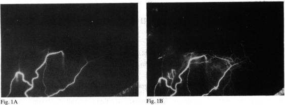
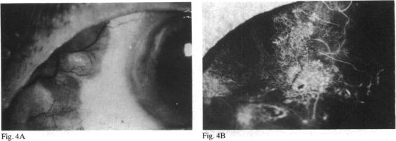
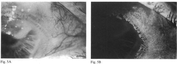
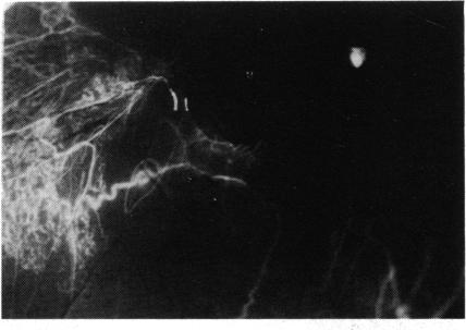
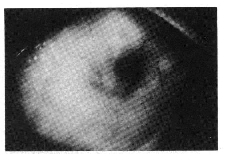
Anterior segment fluorescein angiography has been used in the investigation of patients with sclerokeratitis. This showed that corneal thinning or destruction was associated with non-perfusion of the episcleral vasculature. The changes arose either as a result of a systemic vasculitis in seropositive individuals or were induced by surgery to the eye. Infiltrative forms of sclerokeratitis were commoner in seronegative patients and were less often associated with vascular shutdown.

摘要

前段荧光素血管造影已用于巩膜角膜炎患者的检查。这表明角膜变薄或破坏与巩膜上血管系统的无灌注有关。这些改变要么是血清反应阳性个体全身性血管炎的结果,要么是眼部手术所致。浸润型巩膜角膜炎在血清反应阴性患者中更为常见,且较少与血管关闭相关。

https://cdn.ncbi.nlm.nih.gov/pmc/blobs/a4ef/1041105/494744938818/brjopthal00612-0075-b.jpg
https://cdn.ncbi.nlm.nih.gov/pmc/blobs/a4ef/1041105/5fd269857659/brjopthal00612-0071-a.jpg
https://cdn.ncbi.nlm.nih.gov/pmc/blobs/a4ef/1041105/f78e6c201b1a/brjopthal00612-0071-b.jpg
https://cdn.ncbi.nlm.nih.gov/pmc/blobs/a4ef/1041105/58098359381c/brjopthal00612-0071-c.jpg
https://cdn.ncbi.nlm.nih.gov/pmc/blobs/a4ef/1041105/39cc3c1da569/brjopthal00612-0072-a.jpg
https://cdn.ncbi.nlm.nih.gov/pmc/blobs/a4ef/1041105/d41610238f27/brjopthal00612-0072-b.jpg
https://cdn.ncbi.nlm.nih.gov/pmc/blobs/a4ef/1041105/8ba1ce0eaf4b/brjopthal00612-0073-a.jpg
https://cdn.ncbi.nlm.nih.gov/pmc/blobs/a4ef/1041105/ff486d070a3b/brjopthal00612-0073-b.jpg
https://cdn.ncbi.nlm.nih.gov/pmc/blobs/a4ef/1041105/7e35f219fac2/brjopthal00612-0074-a.jpg
https://cdn.ncbi.nlm.nih.gov/pmc/blobs/a4ef/1041105/552c9d8e8561/brjopthal00612-0074-b.jpg
https://cdn.ncbi.nlm.nih.gov/pmc/blobs/a4ef/1041105/0830f78c699e/brjopthal00612-0075-a.jpg
https://cdn.ncbi.nlm.nih.gov/pmc/blobs/a4ef/1041105/494744938818/brjopthal00612-0075-b.jpg

相似文献

1
Fluorescein angiography in the differential diagnosis of sclerokeratitis.荧光素血管造影在硬化性角膜炎鉴别诊断中的应用
Br J Ophthalmol. 1987 Feb;71(2):145-51. doi: 10.1136/bjo.71.2.145.
2
Anterior segment fluorescein angiography in the diagnosis of scleral inflammation.眼前节荧光血管造影在巩膜炎症诊断中的应用
Ophthalmology. 1985 Jan;92(1):1-11. doi: 10.1016/s0161-6420(85)34074-5.
3
Acanthamoeba sclerokeratitis. Determining diagnostic criteria.
Arch Ophthalmol. 1986 Sep;104(9):1313-7. doi: 10.1001/archopht.1986.01050210067027.
4
[Necrotizing sclerokeratitis following cataract extraction].
Klin Monbl Augenheilkd. 1988 Dec;193(6):642-4. doi: 10.1055/s-2008-1050312.
5
Clinical manifestations and outcome of tuberculous sclerokeratitis.结核性硬化性角膜炎的临床表现及预后
Br J Ophthalmol. 2016 Sep;100(9):1301-3. doi: 10.1136/bjophthalmol-2015-307599. Epub 2015 Dec 23.
6
Anterior segment fluorescein angiography in inflammatory diseases of the cornea.角膜炎症性疾病的眼前节荧光素血管造影
Acta Ophthalmol (Copenh). 1979 Oct;57(5):781-93. doi: 10.1111/j.1755-3768.1979.tb01844.x.
7
Necrotizing sclerokeratitis following uncomplicated cataract surgery.单纯性白内障手术后发生的坏死性巩膜角膜炎。
Acta Ophthalmol Scand. 1998 Jun;76(3):382-3. doi: 10.1034/j.1600-0420.1998.760330.x.
8
Anterior segment fluorescein angiography in the surgery of immunologically induced corneal and scleral destructive disorders.眼前节荧光素血管造影在免疫性诱导的角膜和巩膜破坏性疾病手术中的应用
Ophthalmology. 1987 Nov;94(11):1452-64. doi: 10.1016/s0161-6420(87)33276-2.
9
Fluorescein angiography of the globe and anterior segment.眼球及眼前节荧光素血管造影。
Trans Ophthalmol Soc U K (1962). 1970;90:339-67.
10
[Postoperative necrotizing sclerokeratitis].
Klin Monbl Augenheilkd. 1990 Dec;197(6):503-5. doi: 10.1055/s-2008-1046319.

引用本文的文献

1
Granulomatosis with polyangiitis (Wegener's disease): An updated review of ocular disease manifestations.肉芽肿性多血管炎(韦格纳肉芽肿病):眼部疾病表现的最新综述
Intractable Rare Dis Res. 2016 May;5(2):61-9. doi: 10.5582/irdr.2016.01014.
2
Anterior Segment Scleral Fluorescein Angiography in the Evaluation of Ciliary Body Neoplasm: Two Case Reports.眼前节巩膜荧光素血管造影在睫状体肿瘤评估中的应用:两例报告
Case Rep Ophthalmol. 2016 Jan 8;7(1):30-8. doi: 10.1159/000443603. eCollection 2016 Jan-Apr.
3
Diagnosis and management of systemic Wegener's granulomatosis presenting with anterior ocular inflammatory disease.

本文引用的文献

1
Anterior segment fluorescein angiography in the diagnosis of scleral inflammation.眼前节荧光血管造影在巩膜炎症诊断中的应用
Ophthalmology. 1985 Jan;92(1):1-11. doi: 10.1016/s0161-6420(85)34074-5.
Br J Ophthalmol. 1991 Apr;75(4):201-7. doi: 10.1136/bjo.75.4.201.