Bennett Maureen P, Silver Gena, Tromblee Tonya, Kohler Rickard, Frem Daniel, Glass Eric N, Kent Marc
Section of Neurology and Neurosurgery, Massachusetts Veterinary Referral Hospital, Woburn, MA, United States.
Section of Diagnostic Imaging, Massachusetts Veterinary Referral Hospital, Woburn, MA, United States.
Front Vet Sci. 2024 Jan 24;10:1294395. doi: 10.3389/fvets.2023.1294395. eCollection 2023.
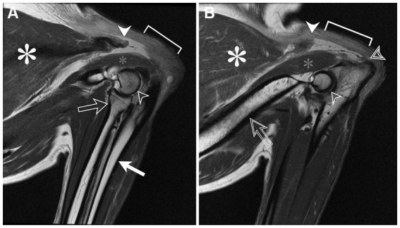
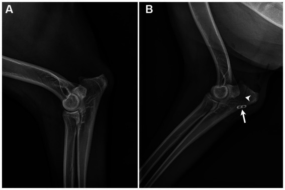
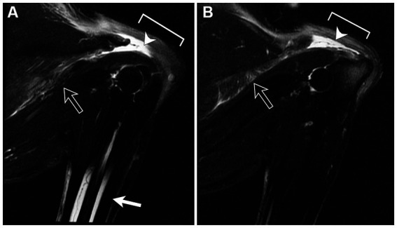
A 7-year-old female spayed Australian shepherd dog was presented for an acute onset of inability to stand. On physical examination, the dog was unable to support weight on the thoracic limbs. On neurological examination, the thoracic limbs had absent hopping and paw placement and reduced withdrawal reflexes bilaterally. The remainder of the neurological examination was normal. The anatomic lesion localized to the C6-T2 spinal nerve roots, spinal nerves, or the named nerves of the thoracic limb, bilaterally. A lesion affecting the ventral gray column of the C6 through T2 spinal cord segments was considered less likely. In an effort to exclude an orthopedic disorder from consideration, radiographs of the shoulders, elbows, and manus were normal. Magnetic resonance imaging of the cervical and cranial thoracic vertebral column was normal. Analysis of synovial fluid from the carpi, elbows, and shoulders were normal. Ultrasonography of the triceps muscle and tendon of insertion revealed bilateral, acute-subacute tears of the tendon at insertion of the triceps muscles, bilaterally. Magnetic resonance imaging of both elbows revealed complete avulsion of the triceps tendons bilaterally. Surgical repair of both tendons was performed using the Arthrex FiberLoop system combined with autologous conditioned plasma soaked in a collagen sponge. Postoperatively, external coaptation was provided using Spica splints for 6 weeks followed by the use of soft padded orthotic braces for an additional 6 weeks. Concurrently, a front support wheelchair was used for 10 weeks postoperative. By 10 weeks postoperative, the dog was able to ambulate without support. To the authors' knowledge, this is the first report of bilateral triceps tendon avulsion in a dog. Tendon avulsion occurred without a known history of trauma or predisposing metabolic abnormalities. Magnetic resonance imaging provided excellent anatomical detail that aided in surgical repair.
一只7岁已绝育的澳大利亚牧羊犬因急性起病无法站立前来就诊。体格检查时,该犬无法用胸肢支撑体重。神经学检查发现,胸肢双侧跳跃和爪放置动作消失,退缩反射减弱。神经学检查的其余部分正常。解剖学病变定位于双侧C6 - T2脊髓神经根、脊神经或胸肢的特定神经。影响C6至T2脊髓节段腹侧灰质柱的病变可能性较小。为排除骨科疾病,肩部、肘部和手部的X线片正常。颈椎和胸段上段脊柱的磁共振成像正常。腕关节、肘关节和肩关节的滑液分析正常。肱三头肌及其止点肌腱的超声检查显示双侧肱三头肌止点肌腱急性 - 亚急性撕裂。双侧肘关节的磁共振成像显示肱三头肌腱完全撕脱。使用Arthrex FiberLoop系统结合浸泡在胶原海绵中的自体条件血浆对双侧肌腱进行手术修复。术后,使用人字形夹板进行外部固定6周,随后再使用软衬垫矫形支具6周。同时,术后使用前支撑轮椅10周。术后10周时,该犬能够在无支撑的情况下行走。据作者所知,这是犬双侧肱三头肌腱撕脱的首例报告。肌腱撕脱发生时无已知的创伤史或易患的代谢异常。磁共振成像提供了有助于手术修复的出色解剖细节。