• 文献检索
  • 文档翻译
  • 深度研究
  • 学术资讯
  • Suppr Zotero 插件Zotero 插件
  • 邀请有礼
  • 套餐&价格
  • 历史记录
应用&插件
Suppr Zotero 插件Zotero 插件浏览器插件Mac 客户端Windows 客户端微信小程序
定价
高级版会员购买积分包购买API积分包
服务
文献检索文档翻译深度研究API 文档MCP 服务
关于我们
关于 Suppr公司介绍联系我们用户协议隐私条款
关注我们

Suppr 超能文献

核心技术专利:CN118964589B侵权必究
粤ICP备2023148730 号-1Suppr @ 2026

文献检索

告别复杂PubMed语法,用中文像聊天一样搜索,搜遍4000万医学文献。AI智能推荐,让科研检索更轻松。

立即免费搜索

文件翻译

保留排版,准确专业,支持PDF/Word/PPT等文件格式,支持 12+语言互译。

免费翻译文档

深度研究

AI帮你快速写综述,25分钟生成高质量综述,智能提取关键信息,辅助科研写作。

立即免费体验

下颌舌侧窝的定量和定性 3D 分析:对下颌后牙区种植规划的影响。

Quantitative and qualitative 3D analysis of mandibular lingual concavities: Implications for dental implant planning in the posterior mandible.

机构信息

Substitutive Dental Science Department, College of Dentistry, Taibah University, Al-Madinah, Saudi Arabia.

Department of Prosthodontics, Faculty of Dentistry, Ibb University, Ibb, Yemen.

出版信息

Clin Exp Dent Res. 2024 Feb;10(1):e858. doi: 10.1002/cre2.858.

DOI:10.1002/cre2.858
PMID:38345362
原文链接:https://pmc.ncbi.nlm.nih.gov/articles/PMC10860544/
Abstract

OBJECTIVE

The purpose of this study is to investigate the type of ridge (degree of angulation of the lingual concavity) and the buccolingual dimensions in the area of the first and second molars in both genders of different ages and how this will affect implant placement in the posterior mandible.

MATERIALS AND METHODS

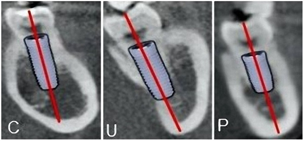
This retrospective cross-sectional study comprised cone beam computed tomography images of 150 dental patients (75 males and 71 aged ≥30). The following were measured/reported: type (morphology) of the ridge (convex [C], parallel [P], or undercut [U]), buccolingual width at the base and the crest of the ridge, and ridge height. The concavity angle, depth, and length of the U-shaped ridge were measured too.

RESULTS

The prevalence of type U ridge ranged from 32.7% in the first molar region to 62.7% in the second molar region. Almost all measurements and ridge type distributions were comparable amongst the age groups (p > .05). Very few significant differences were found when comparing #36 versus #37 and #46 versus #47 teeth, with no differences in the distribution of the ridge types (p > .05). Quite the inverse, all measurements were statistically different when comparing #36 versus #37 and #46 versus #47 teeth, and type U ridge was more frequent in second molar compared to the first molar regions, respectively (p < .05). Many measurements were statistically higher in females; the inverse was true for a few measurements (p < .05). Type U ridge in #36 and #37 was found more frequently among males (p < .001). In contrast, the ridge types in #37 and #47 were not statistically different gender-wise.

CONCLUSIONS

The U type of ridge was more prevalent in the investigated population, encountered more frequently in the second molars generally and in the first molars of males than females. Most posterior mandibular measurements are similar age- and side-wise but seem different gender- and tooth-wise.

摘要

目的

本研究旨在探讨不同性别、不同年龄患者第一、二磨牙区颊舌向宽度和牙槽嵴形态(颊舌向凹陷角度),及其对下颌后牙区种植体植入的影响。

材料和方法

本回顾性横断面研究纳入了 150 例(75 名男性,71 名年龄≥30 岁)患者的锥形束 CT 图像。报告并测量了以下内容:牙槽嵴形态(凸形[C]、平行形[P]或凹陷形[U])、牙槽嵴底部和顶部的颊舌向宽度、牙槽嵴高度。还测量了 U 形牙槽嵴的凹陷角度、深度和长度。

结果

U 形牙槽嵴的发生率从第一磨牙区的 32.7%到第二磨牙区的 62.7%不等。各年龄组之间的各项测量值和嵴形分布差异均无统计学意义(p>.05)。比较 #36 和 #37 以及 #46 和 #47 时,仅少数测量值和嵴形分布存在显著差异(p>.05)。相比之下,比较 #36 和 #37 以及 #46 和 #47 时,所有测量值均有统计学差异,且第二磨牙区的 U 形嵴发生率明显高于第一磨牙区(p<.05)。女性的多数测量值更高,少数测量值则相反(p<.05)。男性 #36 和 #37 的 U 形嵴更为常见(p<.001)。相比之下,#37 和 #47 的嵴形在性别间无统计学差异。

结论

在所研究的人群中,U 形嵴更为常见,一般而言,第二磨牙区的 U 形嵴更为常见,且男性的第一磨牙区 U 形嵴发生率高于女性。大多数下颌后牙区的测量值在年龄和侧别上相似,但在性别和牙位上存在差异。

https://cdn.ncbi.nlm.nih.gov/pmc/blobs/3ead/10860544/d1f2e076c034/CRE2-10-e858-g005.jpg
https://cdn.ncbi.nlm.nih.gov/pmc/blobs/3ead/10860544/1fccaee5a7f0/CRE2-10-e858-g004.jpg
https://cdn.ncbi.nlm.nih.gov/pmc/blobs/3ead/10860544/5129c03c9f0d/CRE2-10-e858-g001.jpg
https://cdn.ncbi.nlm.nih.gov/pmc/blobs/3ead/10860544/5e1e4d47ef15/CRE2-10-e858-g003.jpg
https://cdn.ncbi.nlm.nih.gov/pmc/blobs/3ead/10860544/7590a1a769b7/CRE2-10-e858-g002.jpg
https://cdn.ncbi.nlm.nih.gov/pmc/blobs/3ead/10860544/d1f2e076c034/CRE2-10-e858-g005.jpg

相似文献

1
Quantitative and qualitative 3D analysis of mandibular lingual concavities: Implications for dental implant planning in the posterior mandible.下颌舌侧窝的定量和定性 3D 分析:对下颌后牙区种植规划的影响。
Clin Exp Dent Res. 2024 Feb;10(1):e858. doi: 10.1002/cre2.858.
2
Evaluation of anatomic variations of mandibular lingual concavities from cone beam computed tomography scans in a Malaysian population.评价马来西亚人群下颌舌侧窝的锥形束 CT 扫描解剖变异。
J Prosthet Dent. 2021 May;125(5):766.e1-766.e8. doi: 10.1016/j.prosdent.2021.02.018. Epub 2021 Mar 19.
3
Risk assessment of inferior alveolar nerve injury for immediate implant placement in the posterior mandible: a virtual implant placement study.下颌后牙区即刻种植时下牙槽神经损伤的风险评估:一项虚拟种植体植入研究
J Dent. 2014 Mar;42(3):263-70. doi: 10.1016/j.jdent.2013.12.014. Epub 2014 Jan 3.
4
Cross-sectional analysis of the mandibular lingual concavity using cone beam computed tomography.应用锥形束 CT 对下颌舌侧窝进行的横断面分析。
Clin Oral Implants Res. 2011 Feb;22(2):201-6. doi: 10.1111/j.1600-0501.2010.02018.x. Epub 2010 Oct 6.
5
Effects of Crest Morphology on Lingual Concavity in Mandibular Molar Region: an Observational Study.嵴形态对下颌磨牙区舌侧凹陷的影响:一项观察性研究。
J Oral Maxillofac Res. 2023 Mar 31;14(1):e3. doi: 10.5037/jomr.2023.14103. eCollection 2023 Jan-Mar.
6
Assessment of Lingual Concavities in Submandibular Fossa Region in Patients requiring Dental Implants-A Cone Beam Computed Tomography Study.牙种植患者下颌下窝区域舌侧凹陷的评估——一项锥形束计算机断层扫描研究
J Contemp Dent Pract. 2018 Nov 1;19(11):1329-1333.
7
Radiomorphometric analysis of edentulous posterior mandibular ridges in the first molar region: a cone-beam computed tomography study.第一磨牙区无牙下颌后牙嵴的放射形态计量分析:一项锥形束计算机断层扫描研究。
J Periodontal Implant Sci. 2019 Nov 11;50(1):28-37. doi: 10.5051/jpis.2020.50.1.28. eCollection 2020 Feb.
8
Relationship between CBCT and panoramic images of the morphology and angulation of the posterior mandibular jaw bone.锥形束计算机断层扫描(CBCT)与全景图像在下颌后牙区颌骨形态及角度方面的关系。
Surg Radiol Anat. 2016 Apr;38(3):313-20. doi: 10.1007/s00276-015-1553-1. Epub 2015 Sep 14.
9
Risk assessment of lingual plate perforation in posterior mandibular region: a virtual implant placement study using cone-beam computed tomography.下颌后区舌板穿孔风险评估:锥形束 CT 引导下的虚拟种植体放置研究。
J Periodontol. 2011 Jan;82(1):129-35. doi: 10.1902/jop.2010.100313. Epub 2010 Jul 27.
10
Relevant factors of posterior mandible lingual plate perforation during immediate implant placement: a virtual implant placement study using CBCT.即刻种植时下颌舌板穿孔的相关因素:CBCT 引导下的虚拟种植体放置研究。
BMC Oral Health. 2023 Feb 6;23(1):76. doi: 10.1186/s12903-022-02696-z.

引用本文的文献

1
The prevalence of mandibular lingual concavity among the Saudi population of Eastern Province: A CBCT evaluation.沙特东部省人群下颌舌侧凹陷的患病率:一项锥形束计算机断层扫描评估。
Saudi Dent J. 2024 Dec;36(12):1618-1622. doi: 10.1016/j.sdentj.2024.11.015. Epub 2024 Dec 5.
2
Anatomical changes in alveolar bone structure following the extraction of the mandibular second molar using CBCT measurements.使用锥形束计算机断层扫描(CBCT)测量下颌第二磨牙拔除后牙槽骨结构的解剖学变化。
Sci Rep. 2025 May 22;15(1):17789. doi: 10.1038/s41598-025-02705-5.
3
Evaluation of submandibular fossa depth and mandibular canal relationship with cone-beam computed tomography.

本文引用的文献

1
Relevant factors of posterior mandible lingual plate perforation during immediate implant placement: a virtual implant placement study using CBCT.即刻种植时下颌舌板穿孔的相关因素:CBCT 引导下的虚拟种植体放置研究。
BMC Oral Health. 2023 Feb 6;23(1):76. doi: 10.1186/s12903-022-02696-z.
2
Morphometric Analysis of the Midline Mandibular Lingual Canal and Mandibular Lingual Foramina: A Cone Beam Computed Tomography (CBCT) Evaluation.下颌舌侧管和下颌舌侧孔的形态分析:锥形束计算机断层扫描(CBCT)评估。
Int J Environ Res Public Health. 2022 Dec 16;19(24):16910. doi: 10.3390/ijerph192416910.
3
Influence of bone anatomical morphology of mandibular molars on dental implant based on CBCT.
锥形束计算机断层扫描评估下颌下窝深度及下颌管关系
BMC Oral Health. 2025 Apr 18;25(1):601. doi: 10.1186/s12903-025-05686-z.
4
A Comprehensive Analysis of the Radiographic Characteristics and Bilateral Symmetry of the Mental Foramen.颏孔的影像学特征及双侧对称性的综合分析
Clin Exp Dent Res. 2025 Feb;11(1):e70081. doi: 10.1002/cre2.70081.
5
Anatomic Variations Important for Dental Implantation in the Mandible-A Systematic Review.对下颌骨牙种植重要的解剖变异——一项系统评价
Diagnostics (Basel). 2025 Jan 11;15(2):155. doi: 10.3390/diagnostics15020155.
6
Factors influencing submandibular fossa visibility on panoramic images: a comparative CBCT study.全景影像上下颌下窝可见性的影响因素:一项CBCT对比研究
BMC Oral Health. 2024 Dec 26;24(1):1556. doi: 10.1186/s12903-024-05364-6.
7
The mylohyoid line is highly variable but does not affect the microarchitecture of the edentulous alveolar bone - an anatomical micro-CT study.下颌舌骨线高度可变,但不影响无牙颌牙槽骨的微观结构——一项解剖学显微CT研究
BMC Oral Health. 2024 May 3;24(1):528. doi: 10.1186/s12903-024-04293-8.
基于 CBCT 的下颌磨牙骨解剖形态对牙种植体的影响。
BMC Oral Health. 2021 Oct 15;21(1):528. doi: 10.1186/s12903-021-01888-3.
4
Anatomic considerations for immediate implant placement in the mandibular molar region: a cross-sectional study using cone-beam computed tomography.下颌磨牙区即刻种植的解剖学考虑:使用锥形束 CT 的横断面研究。
Folia Morphol (Warsz). 2022;81(3):732-738. doi: 10.5603/FM.a2021.0060. Epub 2021 Jun 29.
5
Evaluation of anatomic variations of mandibular lingual concavities from cone beam computed tomography scans in a Malaysian population.评价马来西亚人群下颌舌侧窝的锥形束 CT 扫描解剖变异。
J Prosthet Dent. 2021 May;125(5):766.e1-766.e8. doi: 10.1016/j.prosdent.2021.02.018. Epub 2021 Mar 19.
6
Assessing the frequency of deep lingual concavities in 826 posterior mandible sockets.评估 826 个下颌后窝中深舌凹的频率。
J Craniomaxillofac Surg. 2020 Nov;48(11):1045-1051. doi: 10.1016/j.jcms.2020.09.001. Epub 2020 Sep 12.
7
Computed Tomography for the Assessment of the Potential Risk After Implant Placement in Fresh Extraction Sites in the Posterior Mandible.计算机断层扫描评估下颌后牙新鲜拔牙窝内种植体植入后的潜在风险。
J Oral Implantol. 2021 Feb 1;47(1):2-8. doi: 10.1563/aaid-joi-D-18-00227.
8
Radiomorphometric analysis of edentulous posterior mandibular ridges in the first molar region: a cone-beam computed tomography study.第一磨牙区无牙下颌后牙嵴的放射形态计量分析:一项锥形束计算机断层扫描研究。
J Periodontal Implant Sci. 2019 Nov 11;50(1):28-37. doi: 10.5051/jpis.2020.50.1.28. eCollection 2020 Feb.
9
Mandibular residual ridge height in relation to age, gender and duration of edentulism in a Saudi population: A clinical and radiographic study.沙特人群中下颌剩余牙槽嵴高度与年龄、性别及无牙时间的关系:一项临床及影像学研究。
Saudi Dent J. 2019 Apr;31(2):258-264. doi: 10.1016/j.sdentj.2018.12.006. Epub 2019 Jan 8.
10
Comparison of digital panoramic radiography versus cone beam computerized tomography for measuring alveolar bone.数字全景X线摄影术与锥形束计算机断层扫描术在测量牙槽骨方面的比较。
Head Face Med. 2017 Feb 22;13(1):2. doi: 10.1186/s13005-017-0135-3.