Lee Dagyeong, Ham In-Hye, Oh Hye Jeong, Lee Dong Min, Yoon Jung Hwan, Son Sang-Yong, Kim Tae-Min, Kim Jae-Young, Han Sang-Uk, Hur Hoon
Department of Surgery, Ajou University School of Medicine, Suwon, Republic of Korea.
Cancer Biology Graduate Program, Ajou University School of Medicine Suwon, Suwon, Republic of Korea.
J Transl Med. 2024 Feb 14;22(1):154. doi: 10.1186/s12967-024-04963-9.
Tumor cells of diffuse-type gastric cancer (DGC) are discohesive and infiltrate into the stroma as single cells or small subgroups, so the stroma significantly impacts DGC progression. Cancer-associated fibroblasts (CAFs) are major components of the tumor stroma. Here, we identified CAF-specific secreted molecules and investigated the mechanism underlying CAF-induced DGC progression.
We conducted transcriptome analysis for paired normal fibroblast (NF)-CAF isolated from DGC patient tissues and proteomics for conditioned media (CM) of fibroblasts. The effects of fibroblasts on cancer cells were examined by transwell migration and soft agar assays, western blotting, and in vivo. We confirmed the effect of blocking tubulointerstitial nephritis antigen-like 1 (TINAGL1) in CAFs using siRNA or shRNA. We evaluated the expression of TINAGL1 protein in frozen tissues of DGC and paired normal stomach and mRNA in formalin-fixed, paraffin-embedded (FFPE) tissue using RNA in-situ hybridization (RNA-ISH).
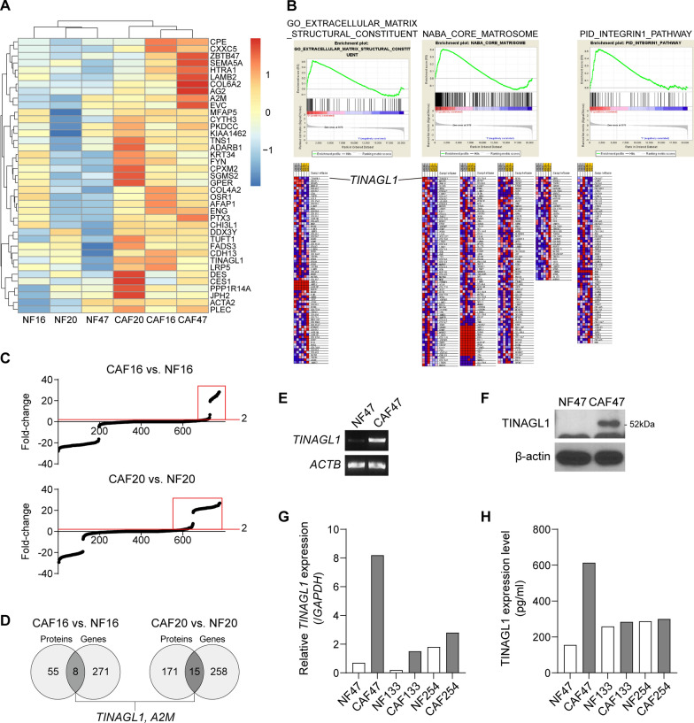
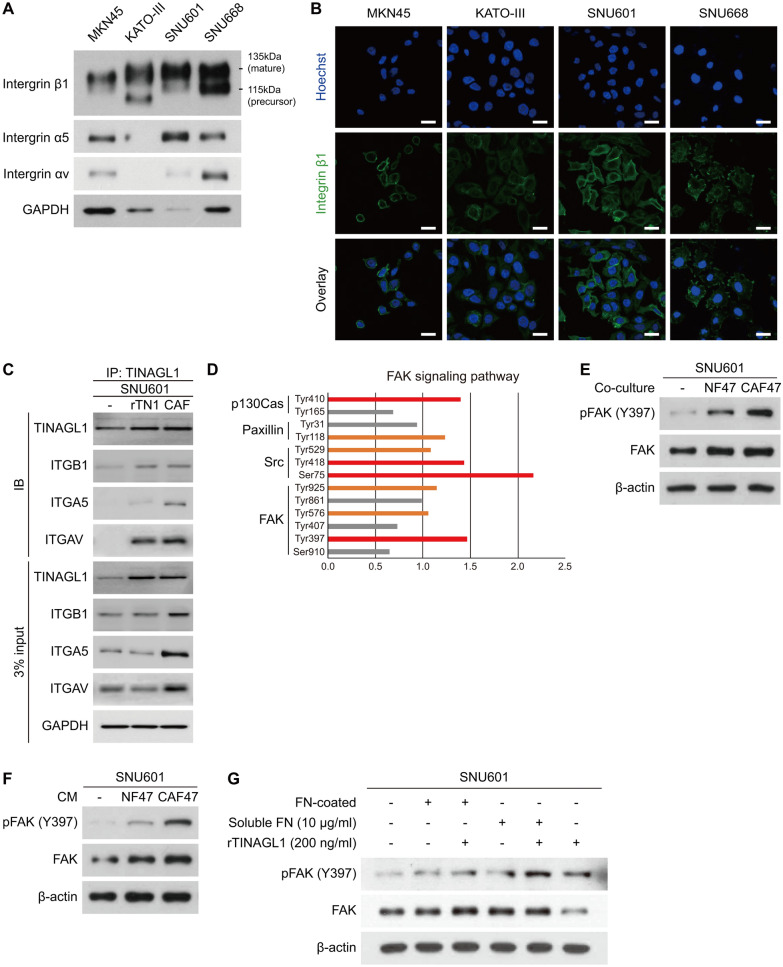
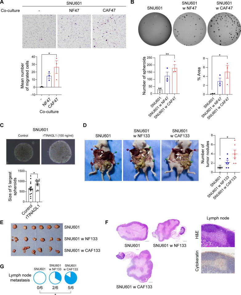
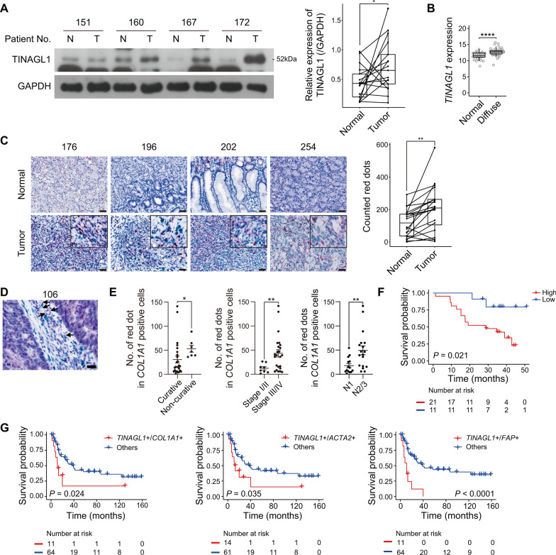
CAFs more highly expressed TINAGL1 than NFs. The co-culture of CAFs increased migration and tumorigenesis of DGC. Moreover, CAFs enhanced the phosphorylation of focal adhesion kinase (FAK) and mesenchymal marker expression in DGC cells. In an animal study, DGC tumors co-injected with CAFs showed aggressive phenotypes, including lymph node metastasis. However, increased phosphorylation of FAK and migration were reduced by blocking TINAGL1 in CAFs. In the tissues of DGC patients, TINAGL1 was higher in cancer than paired normal tissues and detected with collagen type I alpha 1 chain (COL1A1) in the same spot. Furthermore, high TINAGL1 expression was significantly correlated with poor prognosis in several public databases and our patient cohort diagnosed with DGC.
These results indicate that TINAGL1 secreted by CAFs induces phosphorylation of FAK in DGC cells and promotes tumor progression. Thus, targeting TINAGL1 in CAFs can be a novel therapeutic strategy for DGC.
弥漫型胃癌(DGC)的肿瘤细胞缺乏黏附性,以单个细胞或小细胞亚群的形式浸润到基质中,因此基质对DGC的进展有显著影响。癌症相关成纤维细胞(CAF)是肿瘤基质的主要成分。在此,我们鉴定了CAF特异性分泌分子,并研究了CAF诱导DGC进展的机制。
我们对从DGC患者组织中分离出的配对正常成纤维细胞(NF)-CAF进行了转录组分析,并对成纤维细胞的条件培养基(CM)进行了蛋白质组学分析。通过Transwell迁移和软琼脂试验、蛋白质免疫印迹法以及体内实验,研究了成纤维细胞对癌细胞的影响。我们使用小干扰RNA(siRNA)或短发夹RNA(shRNA)证实了在CAF中阻断肾小管间质性肾炎抗原样1(TINAGL1)的作用。我们使用RNA原位杂交(RNA-ISH)评估了DGC冰冻组织及配对正常胃组织中TINAGL1蛋白的表达,以及福尔马林固定、石蜡包埋(FFPE)组织中TINAGL1的mRNA表达。
CAF比NF更高水平地表达TINAGL1。CAF的共培养增加了DGC的迁移和肿瘤发生。此外,CAF增强了DGC细胞中粘着斑激酶(FAK)的磷酸化和间充质标志物的表达。在一项动物研究中,与CAF共同注射的DGC肿瘤表现出侵袭性表型,包括淋巴结转移。然而,通过在CAF中阻断TINAGL1,可降低FAK磷酸化增加和迁移。在DGC患者的组织中,癌组织中TINAGL1高于配对的正常组织,并且在同一部位与I型胶原α1链(COL1A1)共同检测到。此外,在几个公共数据库以及我们诊断为DGC的患者队列中,高TINAGL1表达与不良预后显著相关。
这些结果表明,CAF分泌的TINAGL1诱导DGC细胞中FAK的磷酸化并促进肿瘤进展。因此,靶向CAF中的TINAGL1可能是DGC的一种新型治疗策略。