• 文献检索
  • 文档翻译
  • 深度研究
  • 学术资讯
  • Suppr Zotero 插件Zotero 插件
  • 邀请有礼
  • 套餐&价格
  • 历史记录
应用&插件
Suppr Zotero 插件Zotero 插件浏览器插件Mac 客户端Windows 客户端微信小程序
定价
高级版会员购买积分包购买API积分包
服务
文献检索文档翻译深度研究API 文档MCP 服务
关于我们
关于 Suppr公司介绍联系我们用户协议隐私条款
关注我们

Suppr 超能文献

核心技术专利:CN118964589B侵权必究
粤ICP备2023148730 号-1Suppr @ 2026

文献检索

告别复杂PubMed语法,用中文像聊天一样搜索,搜遍4000万医学文献。AI智能推荐,让科研检索更轻松。

立即免费搜索

文件翻译

保留排版,准确专业,支持PDF/Word/PPT等文件格式,支持 12+语言互译。

免费翻译文档

深度研究

AI帮你快速写综述,25分钟生成高质量综述,智能提取关键信息,辅助科研写作。

立即免费体验

第三脑室的立体定向解剖。

Stereotactic anatomy of the third ventricle.

机构信息

Unit of Anatomy, Department of Biomedicine, Faculty of Medicine, University of Porto, Alameda Professor Hernâni Monteiro, 4200‑319, Porto, Portugal.

CINTESIS@RISE, Rua Dr. Plácido da Costa, s/n, 4200‑450, Porto, Portugal.

出版信息

Surg Radiol Anat. 2024 Mar;46(3):271-283. doi: 10.1007/s00276-024-03312-1. Epub 2024 Feb 19.

DOI:10.1007/s00276-024-03312-1
PMID:38374441
原文链接:https://pmc.ncbi.nlm.nih.gov/articles/PMC10960742/
Abstract

PURPOSE

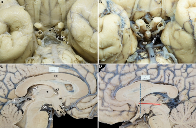
Endoscopic third ventriculostomy (ETV) is a surgical procedure that can lead to complications and requires detailed preoperative planning. This study aimed to provide a more accurate understanding of the anatomy of the third ventricle and the location of important structures to improve the safety and success of ETV.

METHODS

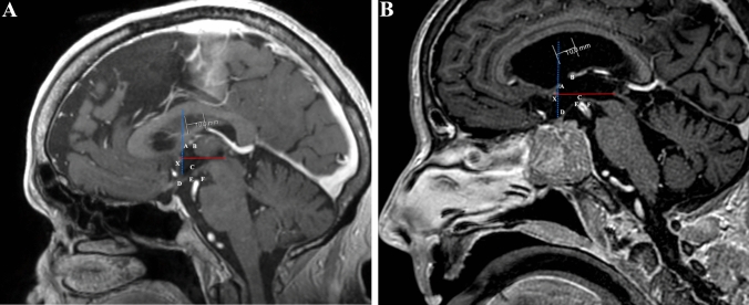
We measured the stereotactic coordinates of six points of interest relative to a predefined stereotactic reference point in 23 cadaver brain hemi-sections, 200 normal brain magnetic resonance imaging (MRI) scans, and 24 hydrocephalic brain MRI scans. The measurements were statistically analyzed, and comparisons were made.

RESULTS

We found some statistically significant differences between genders in MRIs from healthy subjects. We also found statistically significant differences between MRIs from healthy subjects and both cadaver brains and MRIs with hydrocephalus, though their magnitude is very small and not clinically relevant. Some stereotactic points were more posteriorly and inferiorly located in cadaver brains, particularly the infundibular recess and the basilar artery. It was found that all stereotactic points studied were more posteriorly located in brains with hydrocephalus.

CONCLUSION

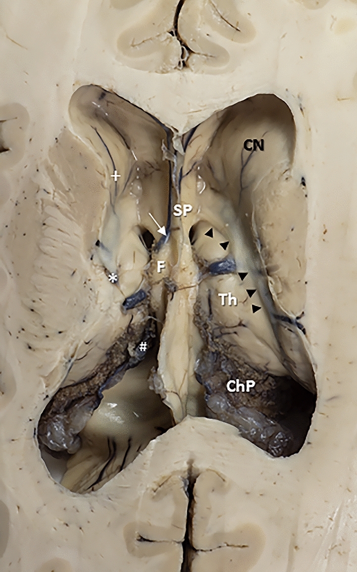
The study describes periventricular structures in cadaver brains and MRI scans from healthy and hydrocephalic subjects, which can guide neurosurgeons in planning surgical approaches to the third ventricle. Overall, the study contributes to understanding ETV and provides insights for improving its safety and efficacy. The findings also support that practicing on cadaveric brains can still provide valuable information and is valid for study and training of neurosurgeons unfamiliar with the ETV technique.

摘要

目的

内镜第三脑室造瘘术(ETV)是一种可能导致并发症的手术,需要详细的术前规划。本研究旨在更准确地了解第三脑室的解剖结构和重要结构的位置,以提高 ETV 的安全性和成功率。

方法

我们在 23 个尸脑半脑切片、200 个正常脑磁共振成像(MRI)扫描和 24 个脑积水脑 MRI 扫描中,测量了六个感兴趣点相对于预定立体定向参考点的立体定向坐标。对测量结果进行了统计学分析,并进行了比较。

结果

我们发现,健康受试者的 MRI 存在一些统计学上的性别差异。我们还发现,健康受试者的 MRI 与尸脑和脑积水 MRI 之间存在统计学上的显著差异,尽管其幅度很小,且无临床意义。在尸脑中,一些立体定向点的位置更靠后和更低,特别是漏斗隐窝和基底动脉。所有研究的立体定向点在脑积水脑内的位置都更靠后。

结论

本研究描述了尸脑和健康及脑积水受试者的 MRI 扫描中的脑室周围结构,可指导神经外科医生规划第三脑室手术入路。总体而言,该研究有助于理解 ETV,并为提高其安全性和有效性提供了思路。研究结果还表明,在不熟悉 ETV 技术的神经外科医生中,在尸脑上进行操作仍然可以提供有价值的信息,并且是有效的研究和培训方法。

https://cdn.ncbi.nlm.nih.gov/pmc/blobs/01f5/10960742/e7614dec4e4b/276_2024_3312_Fig3_HTML.jpg
https://cdn.ncbi.nlm.nih.gov/pmc/blobs/01f5/10960742/7521215c806e/276_2024_3312_Fig1_HTML.jpg
https://cdn.ncbi.nlm.nih.gov/pmc/blobs/01f5/10960742/c4d0e29e14f5/276_2024_3312_Fig2_HTML.jpg
https://cdn.ncbi.nlm.nih.gov/pmc/blobs/01f5/10960742/e7614dec4e4b/276_2024_3312_Fig3_HTML.jpg
https://cdn.ncbi.nlm.nih.gov/pmc/blobs/01f5/10960742/7521215c806e/276_2024_3312_Fig1_HTML.jpg
https://cdn.ncbi.nlm.nih.gov/pmc/blobs/01f5/10960742/c4d0e29e14f5/276_2024_3312_Fig2_HTML.jpg
https://cdn.ncbi.nlm.nih.gov/pmc/blobs/01f5/10960742/e7614dec4e4b/276_2024_3312_Fig3_HTML.jpg

相似文献

1
Stereotactic anatomy of the third ventricle.第三脑室的立体定向解剖。
Surg Radiol Anat. 2024 Mar;46(3):271-283. doi: 10.1007/s00276-024-03312-1. Epub 2024 Feb 19.
2
Operative planning aid for optimal endoscopic third ventriculostomy entry points in pediatric cases.小儿病例中优化内镜下第三脑室造瘘术入路点的手术规划辅助工具。
Childs Nerv Syst. 2017 Feb;33(2):269-273. doi: 10.1007/s00381-016-3320-y. Epub 2017 Jan 18.
3
Transependymal Edema as a Predictor of Endoscopic Third Ventriculostomy Success in Pediatric Hydrocephalus.经室管膜水肿作为小儿脑积水内镜下第三脑室造瘘术成功的预测指标
World Neurosurg. 2021 Dec;156:e215-e221. doi: 10.1016/j.wneu.2021.09.031. Epub 2021 Sep 21.
4
Comparative analysis of endoscopic third ventriculostomy trajectories in pediatric cases.小儿病例中内镜下第三脑室造瘘术轨迹的比较分析
J Neurosurg Pediatr. 2015 Dec;16(6):626-32. doi: 10.3171/2015.4.PEDS14430. Epub 2015 Sep 4.
5
Third ventricle floor bowing: a useful measurement to predict endoscopic third ventriculostomy success in infantile hydrocephalus.第三脑室底凹陷:预测婴幼儿脑积水内镜第三脑室造瘘术成功的有用指标。
Acta Neurochir (Wien). 2020 Jan;162(1):31-37. doi: 10.1007/s00701-019-04133-7. Epub 2019 Dec 2.
6
Endoscopic third ventriculostomy and choroid plexus cauterization in infant hydrocephalus: a prospective study by the Hydrocephalus Clinical Research Network.婴儿脑积水的内镜下第三脑室造瘘术和脉络丛烧灼术:脑积水临床研究网络的一项前瞻性研究
J Neurosurg Pediatr. 2018 Mar;21(3):214-223. doi: 10.3171/2017.8.PEDS17217. Epub 2017 Dec 15.
7
Frame-Based Stereotactic Endoscopic Third Ventriculostomy-Toward Improved Precision and Minimizing Morbidities.基于框架的立体定向内镜第三脑室造瘘术——提高精度和降低发病率。
World Neurosurg. 2020 Aug;140:e240-e246. doi: 10.1016/j.wneu.2020.05.040. Epub 2020 May 11.
8
Anatomy of the floor of the third ventricle in relation to endoscopic ventriculostomy.第三脑室底与内镜下脑室造瘘术相关的解剖结构
Clin Anat. 2009 Nov;22(8):916-24. doi: 10.1002/ca.20867.
9
Endoscopic third ventriculostomy in children: prospective, multicenter results from the Hydrocephalus Clinical Research Network.儿童内镜下第三脑室造瘘术:脑积水临床研究网络的前瞻性多中心研究结果
J Neurosurg Pediatr. 2016 Oct;18(4):423-429. doi: 10.3171/2016.4.PEDS163. Epub 2016 Jun 3.
10
Surgical outcome of tuberculous meningitis hydrocephalus treated by endoscopic third ventriculostomy: prognostic factors and postoperative neuroimaging for functional assessment of ventriculostomy.经内镜第三脑室造瘘术治疗结核性脑膜炎脑积水的手术结果:预后因素及用于脑室造瘘功能评估的术后神经影像学表现
J Neurosurg Pediatr. 2009 May;3(5):371-7. doi: 10.3171/2009.1.PEDS0947.

引用本文的文献

1
Optimal trajectory of the neuroendoscope for third ventricle pavement access.经第三脑室底部入路神经内镜的最佳轨迹
Front Neuroanat. 2025 Jan 22;19:1431128. doi: 10.3389/fnana.2025.1431128. eCollection 2025.
2
: An intraoperative image.一张术中图像。
Brain Spine. 2024 Nov 23;4:104144. doi: 10.1016/j.bas.2024.104144. eCollection 2024.

本文引用的文献

1
The Roof of the 4th Ventricle Seen From Inside: Endoscopic Anatomic Illustration-A Case Series.从内部观察第四脑室顶:内镜解剖示意图——病例系列。
Oper Neurosurg (Hagerstown). 2023 Jul 1;25(1):11-19. doi: 10.1227/ons.0000000000000669. Epub 2023 Mar 3.
2
Anatomic variations of the floor of the third ventricle: Surgical implications for endoscopic third ventriculostomy.第三脑室底部的解剖变异:内镜下第三脑室造瘘术的手术意义
Surg Neurol Int. 2022 May 27;13:218. doi: 10.25259/SNI_404_2022. eCollection 2022.
3
Where the central canal begins: endoscopic in vivo description.
中央管起始部位的内镜下活体描述。
J Neurosurg. 2021 Aug 13;136(3):895-904. doi: 10.3171/2020.12.JNS203649. Print 2022 Mar 1.
4
Anatomic Variability of the Morphometric Parameters of the Third Ventricle of the Brain and Its Relations to the Shape of the Skull.脑第三脑室形态计量学参数的解剖变异及其与颅骨形状的关系。
J Neurol Surg B Skull Base. 2021 Jul;82(Suppl 3):e315-e320. doi: 10.1055/s-0040-1701527. Epub 2020 Feb 7.
5
Neurosurgical anatomy of the floor of the third ventricle and related vascular structures.第三脑室底部的神经外科解剖结构及相关血管结构。
Surg Radiol Anat. 2021 Dec;43(12):1915-1925. doi: 10.1007/s00276-021-02785-8. Epub 2021 Jun 14.
6
Endoscopic Third Ventriculostomy: Role of Image Guidance in Reducing the Complications.内镜下第三脑室造瘘术:影像引导在降低并发症中的作用
Asian J Neurosurg. 2020 Oct 19;15(4):926-930. doi: 10.4103/ajns.AJNS_161_20. eCollection 2020 Oct-Dec.
7
Frame-Based Stereotactic Endoscopic Third Ventriculostomy-Toward Improved Precision and Minimizing Morbidities.基于框架的立体定向内镜第三脑室造瘘术——提高精度和降低发病率。
World Neurosurg. 2020 Aug;140:e240-e246. doi: 10.1016/j.wneu.2020.05.040. Epub 2020 May 11.
8
Neurocognitive Complications after Ventricular Neuroendoscopy: A Systematic Review.脑室神经内镜术后的神经认知并发症:系统评价。
Behav Neurol. 2020 Mar 25;2020:2536319. doi: 10.1155/2020/2536319. eCollection 2020.
9
Marking Basilar Artery Using Neuronavigation During Endoscopic Third Ventriculostomy: A Clinical Study.神经内镜下第三脑室造瘘术中使用神经导航标记基底动脉:一项临床研究
Turk Neurosurg. 2020;30(1):23-29. doi: 10.5137/1019-5149.JTN.25698-19.1.
10
Cadaver Dissection Is Obsolete in Medical Training! A Misinterpreted Notion.尸体解剖在医学培训中已经过时了!这是一个被误解的观念。
Med Princ Pract. 2018;27(3):201-210. doi: 10.1159/000488320. Epub 2018 Mar 12.