• 文献检索
  • 文档翻译
  • 深度研究
  • 学术资讯
  • Suppr Zotero 插件Zotero 插件
  • 邀请有礼
  • 套餐&价格
  • 历史记录
应用&插件
Suppr Zotero 插件Zotero 插件浏览器插件Mac 客户端Windows 客户端微信小程序
定价
高级版会员购买积分包购买API积分包
服务
文献检索文档翻译深度研究API 文档MCP 服务
关于我们
关于 Suppr公司介绍联系我们用户协议隐私条款
关注我们

Suppr 超能文献

核心技术专利:CN118964589B侵权必究
粤ICP备2023148730 号-1Suppr @ 2026

文献检索

告别复杂PubMed语法,用中文像聊天一样搜索,搜遍4000万医学文献。AI智能推荐,让科研检索更轻松。

立即免费搜索

文件翻译

保留排版,准确专业,支持PDF/Word/PPT等文件格式,支持 12+语言互译。

免费翻译文档

深度研究

AI帮你快速写综述,25分钟生成高质量综述,智能提取关键信息,辅助科研写作。

立即免费体验

翼状胬肉中的树突状黑素细胞增生:与原发性后天性黑素沉着症诊断混淆的潜在来源。

Dendritic Melanocytic Hyperplasia in Pterygia: A Potential Source of Diagnostic Confusion with Primary Acquired Melanosis.

作者信息

Oh Angela J, Glasgow Ben J

机构信息

Departments of Ophthalmology, Los Angeles, CA, USA.

Pathology and Laboratory Medicine, Jules Stein Eye Institute, University of California, Los Angeles, CA, USA.

出版信息

Ocul Oncol Pathol. 2023 Aug;9(1-2):48-55. doi: 10.1159/000530514. Epub 2023 Apr 7.

DOI:10.1159/000530514
PMID:38376097
原文链接:https://pmc.ncbi.nlm.nih.gov/articles/PMC10821784/
Abstract

INTRODUCTION

The aim of this study was to report the nearly ubiquitous prevalence of melanocytic hyperplasia in benign pterygia/pingueculae and establish that the entity is insufficiently recognized.

METHODS

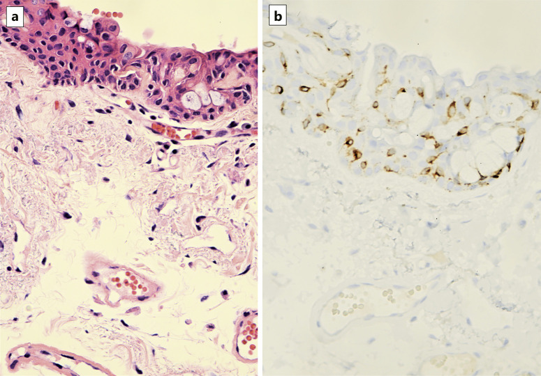
This is a retrospective immunohistochemical pathology case series of 30 consecutive pterygia/pingueculae samples selected from an ophthalmic pathology database at a single institution. Histopathologic and immunohistochemistry analyses with anti-SOX-10 and anti-MART-1 antibodies were used for identifying melanocytes. The number of squamous cells intervening between melanocytes was determined.

RESULTS

The frequency of dendritic melanocytes was found to meet the criteria for dendritic melanocytic hyperplasia in 29 of 30 pterygia/pingueculae samples using specific antibodies. Melanocytes were found in several patterns: diffuse (28%), multifocal (28%), and focal (44%). In each case, the melanocytes were distributed as single melanocytes at the base; clusters of melanocytes were seen in 17% of samples. There were an average of about two intervening epithelial cells between melanocytes at the base.

CONCLUSION

When diagnosed with immunohistochemistry, dendritic melanocytic hyperplasia is nearly ubiquitous in pterygia and pingueculae. Melanocytic hyperplasia may have a distribution that includes nests and single melanocytes above the basal layer, which can be confused with forms of primary acquired melanosis. It is important for pathologists to recognize these lesions as a distinct benign clinicopathologic entity.

摘要

引言

本研究的目的是报告良性翼状胬肉/睑裂斑中黑素细胞增生几乎普遍存在的情况,并确定该实体未得到充分认识。

方法

这是一个回顾性免疫组化病理病例系列,从单一机构的眼科病理数据库中选取了连续30个翼状胬肉/睑裂斑样本。使用抗SOX-10和抗MART-1抗体进行组织病理学和免疫组化分析以识别黑素细胞。确定黑素细胞之间的鳞状细胞数量。

结果

使用特异性抗体,在30个翼状胬肉/睑裂斑样本中的29个中,发现树突状黑素细胞的频率符合树突状黑素细胞增生的标准。黑素细胞有几种分布模式:弥漫性(28%)、多灶性(28%)和局灶性(44%)。在每种情况下,黑素细胞在基底部分布为单个黑素细胞;17%的样本中可见黑素细胞簇。基底处黑素细胞之间平均约有两个间隔的上皮细胞。

结论

通过免疫组化诊断时,树突状黑素细胞增生在翼状胬肉和睑裂斑中几乎普遍存在。黑素细胞增生的分布可能包括基底层上方的巢状和单个黑素细胞,这可能与原发性后天性黑变病的形式相混淆。病理学家认识到这些病变是一种独特的良性临床病理实体很重要。

https://cdn.ncbi.nlm.nih.gov/pmc/blobs/5b28/10821784/c23a2d8ac69c/oop530514_f05.jpg
https://cdn.ncbi.nlm.nih.gov/pmc/blobs/5b28/10821784/1748cebffe49/oop530514_f01.jpg
https://cdn.ncbi.nlm.nih.gov/pmc/blobs/5b28/10821784/25ff2f31db36/oop530514_f02.jpg
https://cdn.ncbi.nlm.nih.gov/pmc/blobs/5b28/10821784/0ab8d4bcca2d/oop530514_f03.jpg
https://cdn.ncbi.nlm.nih.gov/pmc/blobs/5b28/10821784/1059718b9f6c/oop530514_f04.jpg
https://cdn.ncbi.nlm.nih.gov/pmc/blobs/5b28/10821784/c23a2d8ac69c/oop530514_f05.jpg

相似文献

1
Dendritic Melanocytic Hyperplasia in Pterygia: A Potential Source of Diagnostic Confusion with Primary Acquired Melanosis.翼状胬肉中的树突状黑素细胞增生:与原发性后天性黑素沉着症诊断混淆的潜在来源。
Ocul Oncol Pathol. 2023 Aug;9(1-2):48-55. doi: 10.1159/000530514. Epub 2023 Apr 7.
2
The ultrastructure of conjunctival melanocytic tumors.结膜黑素细胞肿瘤的超微结构
Trans Am Ophthalmol Soc. 1984;82:599-752.
3
Conjunctival Primary Acquired Melanosis: Is It Time for a New Terminology?结膜原发性获得性黑素沉着症:是时候采用新术语了吗?
Am J Ophthalmol. 2016 Feb;162:3-19.e1. doi: 10.1016/j.ajo.2015.11.003. Epub 2015 Nov 10.
4
Pigmentation of the Lacrimal Sac Epithelium.泪囊上皮色素沉着。
Ophthalmic Plast Reconstr Surg. 2016 Nov/Dec;32(6):415-423. doi: 10.1097/IOP.0000000000000541.
5
Usefulness of a red chromagen in the diagnosis of melanocytic lesions of the conjunctiva.结膜黑色素细胞病变中红色素的诊断价值。
JAMA Ophthalmol. 2014 May;132(5):622-9. doi: 10.1001/jamaophthalmol.2013.8216.
6
Relationship between Conjunctival Intraepithelial Dendritic Melanocytes and Nevocytes.结膜上皮内树突状黑素细胞与痣细胞的关系。
Ocul Oncol Pathol. 2020 Aug;6(4):251-258. doi: 10.1159/000505492. Epub 2020 Feb 14.
7
Melanosis, melanocytic hyperlasia, and primary malignant melanoma of the nasal cavity.鼻腔黑变病、黑素细胞增生及原发性恶性黑色素瘤
Cancer. 1979 Oct;44(4):1424-33. doi: 10.1002/1097-0142(197910)44:4<1424::aid-cncr2820440438>3.0.co;2-l.
8
[Intraepidermal distribution patterns of melanocytes in the melanocytic lesions on the sole: proposed criteria for histopathologic diagnosis of plantar malignant melanoma in situ].[足底黑素细胞性病变中黑素细胞的表皮内分布模式:原位足底恶性黑素瘤组织病理学诊断的拟议标准]
Nihon Hifuka Gakkai Zasshi. 1989 Apr;99(5):553-60.
9
Comparison of SOX-10, HMB-45, and Melan-A in Benign Melanocytic Lesions.SOX-10、HMB-45和Melan-A在良性黑素细胞性病变中的比较
Clin Cosmet Investig Dermatol. 2021 Oct 5;14:1419-1425. doi: 10.2147/CCID.S333376. eCollection 2021.
10
Clinicopathologic characteristics of premalignant and malignant melanocytic lesions of the conjunctiva.结膜黑色素细胞癌前病变和恶性病变的临床病理特征
Ophthalmology. 1989 Feb;96(2):147-66. doi: 10.1016/s0161-6420(89)32920-4.

本文引用的文献

1
Comparison of SOX-10, HMB-45, and Melan-A in Benign Melanocytic Lesions.SOX-10、HMB-45和Melan-A在良性黑素细胞性病变中的比较
Clin Cosmet Investig Dermatol. 2021 Oct 5;14:1419-1425. doi: 10.2147/CCID.S333376. eCollection 2021.
2
Conjunctival Melanocytic Lesions.结膜黑素细胞病变
Arch Pathol Lab Med. 2022 May 1;146(5):632-646. doi: 10.5858/arpa.2021-0006-RA.
3
Histopathological Variability and Concomitant Lesions in Pterygium in a Large Case Series.大样本病例系列中翼状胬肉的组织病理学变异性及伴随病变
J Ophthalmol. 2021 Mar 19;2021:6623794. doi: 10.1155/2021/6623794. eCollection 2021.
4
Validation of the Newly Proposed World Health Organization Classification System for Conjunctival Melanocytic Intraepithelial Lesions: A Comparison with the C-MIN and PAM Classification Schemes.验证新提出的世界卫生组织结膜黑色素细胞上皮内病变分类系统:与 C-MIN 和 PAM 分类方案的比较。
Am J Ophthalmol. 2021 Mar;223:60-74. doi: 10.1016/j.ajo.2020.10.020. Epub 2020 Oct 30.
5
Immunohistochemical Profiling of Conjunctival Melanocytic Intraepithelial Lesions, Including SOX10, HMB45, Ki67, and P16.结膜黑色素细胞上皮内病变的免疫组织化学特征分析,包括 SOX10、HMB45、Ki67 和 P16。
Am J Ophthalmol. 2021 Feb;222:148-156. doi: 10.1016/j.ajo.2020.09.033. Epub 2020 Sep 28.
6
Conjunctival nevi and melanoma: multiparametric immunohistochemical analysis, including p16, SOX10, HMB45, and Ki-67.结膜痣和黑色素瘤:包括 p16、SOX10、HMB45 和 Ki-67 的多参数免疫组织化学分析。
Hum Pathol. 2020 Sep;103:107-119. doi: 10.1016/j.humpath.2020.07.020. Epub 2020 Jul 21.
7
Naming the Precursors of Conjunctival Melanoma.结膜黑色素瘤的前驱病变命名
Am J Ophthalmol. 2016 Feb;162:1-2. doi: 10.1016/j.ajo.2015.11.037. Epub 2015 Dec 19.
8
Conjunctival Primary Acquired Melanosis: Is It Time for a New Terminology?结膜原发性获得性黑素沉着症:是时候采用新术语了吗?
Am J Ophthalmol. 2016 Feb;162:3-19.e1. doi: 10.1016/j.ajo.2015.11.003. Epub 2015 Nov 10.
9
Differences in tumor thickness between hematoxylin and eosin and Melan-A immunohistochemically stained primary cutaneous melanomas.苏木精和伊红染色与Melan-A免疫组化染色的原发性皮肤黑色素瘤之间肿瘤厚度的差异。
Am J Dermatopathol. 2013 Feb;35(1):56-63. doi: 10.1097/DAD.0b013e31825ba933.
10
Ophthalmic pterygium: a stem cell disorder with premalignant features.眼翼状胬肉:一种具有潜在恶性特征的干细胞疾病。
Am J Pathol. 2011 Feb;178(2):817-27. doi: 10.1016/j.ajpath.2010.10.037.