Department of Pediatrics, Gynecology and Obstetrics, Division of General Pediatrics, Pediatric Hematology and Oncology Unit, University Hospitals of Geneva, Geneva, Switzerland.
Cansearch Research Platform for Pediatric Oncology and Hematology, Faculty of Medicine, Department of Pediatrics, Gynecology and Obstetrics, University of Geneva, Geneva, Switzerland.
Childs Nerv Syst. 2024 Apr;40(4):1053-1064. doi: 10.1007/s00381-023-06272-w. Epub 2024 Feb 20.
Brain stem tumors in children < 3 months at diagnosis are extremely rare. Our aim is to study a retrospective cohort to improve the understanding of the disease course and guide patient management.
This is a multicenter retrospective analysis across the European Society for Pediatric Oncology SIOP-E HGG/DIPG Working Group linked centers, including patients with a brainstem tumor diagnosed between 2009 and 2020 and aged < 3 months at diagnosis. Clinical data were collected, and imaging characteristics were analyzed blindly and independently by two neuroradiologists.
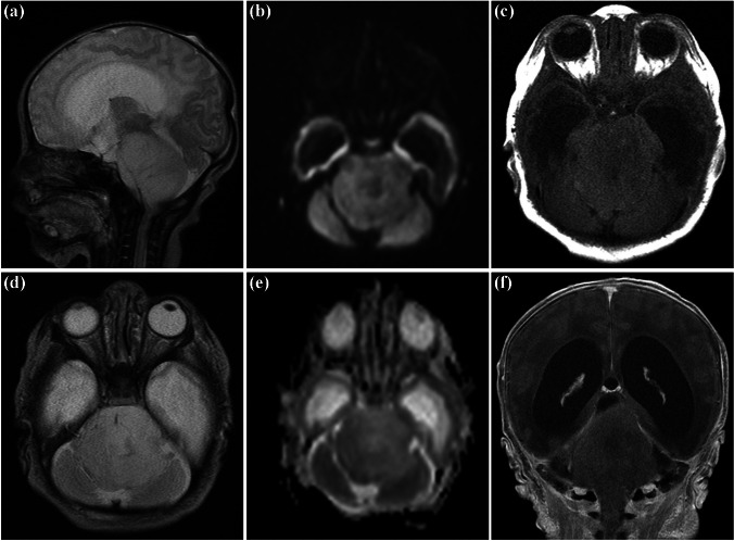
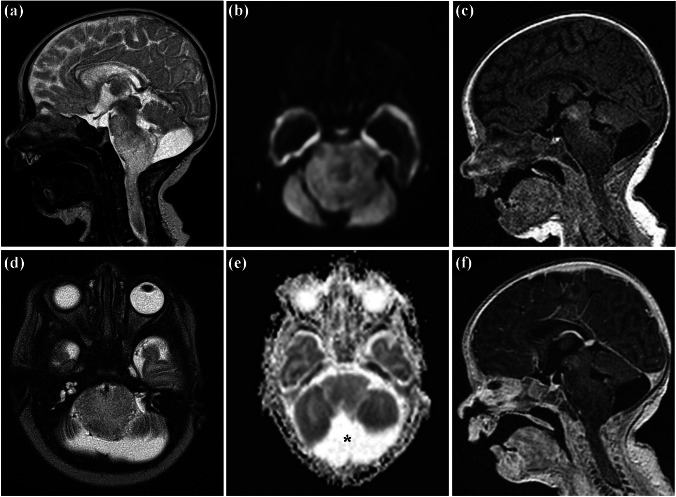
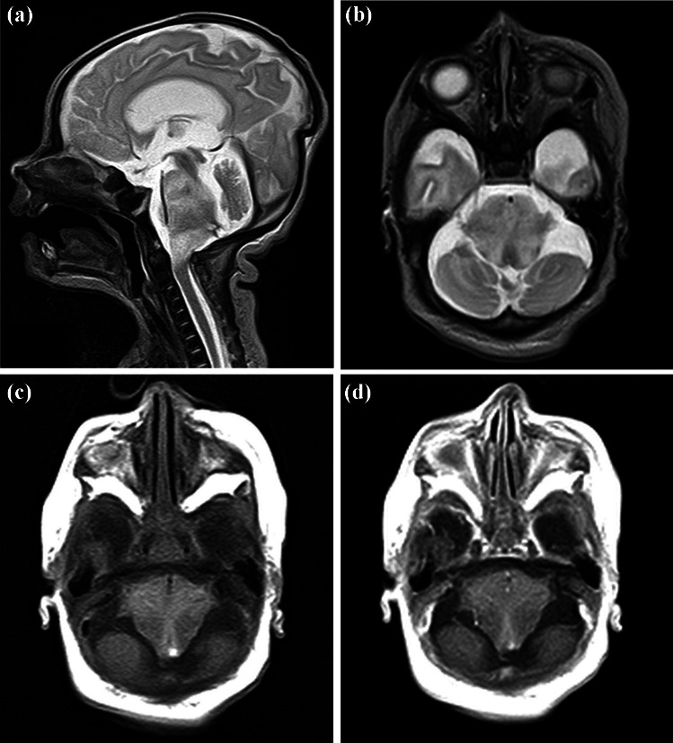
Five cases were identified. No patient received any therapy. The epicenter of two tumors was in the medulla oblongata alone and in the medulla oblongata and the pons in three. For patients with tumor in equal parts in the medulla oblongata and the pons (n = 3), the extension at diagnosis involved the spinal cord; for the two patients with the tumor epicenter in the medulla oblongata alone (n = 2), the extension at diagnosis included the pons (n = 2) and the spinal cord (n = 1). Biopsy was performed in one patient identifying a pilocytic astrocytoma. Two patients died. In one patient, autopsy revealed a high-grade glioma (case 3). Three survivors showed either spontaneous tumor regression (n = 2) or stable disease (n = 1). Survivors were followed up for 10, 7, and 0.6 years, respectively. One case had the typical imaging characteristics of a dorsal exophytic low-grade glioma.
No patient fulfilled the radiologic criteria defining a high-grade glioma. Central neuroradiological review and biopsy may provide useful information regarding the patient management.
3 个月以下诊断为脑干肿瘤的儿童极为罕见。我们旨在研究一个回顾性队列,以提高对疾病过程的认识并指导患者管理。
这是一项跨欧洲儿科肿瘤学会 SIOP-E HGG/DIPG 工作组相关中心的多中心回顾性分析,包括 2009 年至 2020 年间诊断为脑干肿瘤且诊断时年龄<3 个月的患者。收集临床数据,并由两名神经放射科医生对影像学特征进行盲法和独立分析。
共发现 5 例病例。没有患者接受任何治疗。2 例肿瘤的中心位于延髓,3 例位于延髓和脑桥。对于肿瘤延髓和脑桥部分等大的患者(n=3),诊断时的肿瘤延伸累及脊髓;对于肿瘤中心位于延髓的 2 例患者(n=2),诊断时的肿瘤延伸累及脑桥(n=2)和脊髓(n=1)。对 1 例患者进行了活检,确定为毛细胞型星形细胞瘤。2 例患者死亡。1 例患者尸检显示高级别胶质瘤(病例 3)。3 例存活者的肿瘤要么自发消退(n=2),要么病情稳定(n=1)。幸存者的随访时间分别为 10、7 和 0.6 年。1 例患者具有典型的背侧外生低级别胶质瘤影像学特征。
没有患者符合定义高级别胶质瘤的影像学标准。中枢神经影像学复查和活检可能为患者管理提供有用信息。