Department of Microbiology, Tumor and Cell Biology, Karolinska Institutet, Stockholm, Sweden.
Department of Molecular and Clinical Medicine/Wallenberg Laboratory, Institute of Medicine, Sahlgrenska Academy, University of Gothenburg, Gothenburg, Sweden.
Front Immunol. 2024 Feb 7;14:1332733. doi: 10.3389/fimmu.2023.1332733. eCollection 2023.
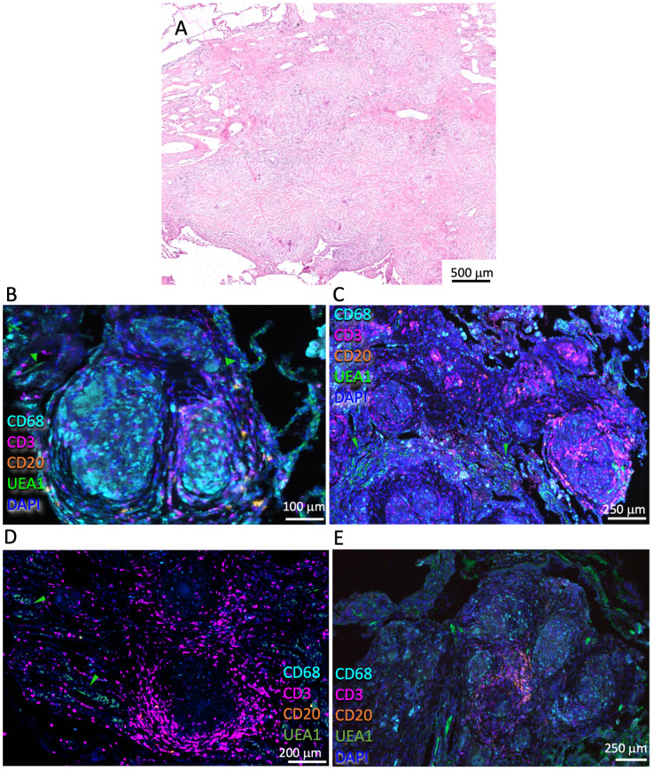
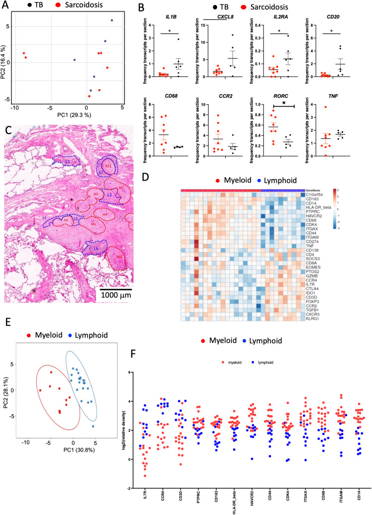
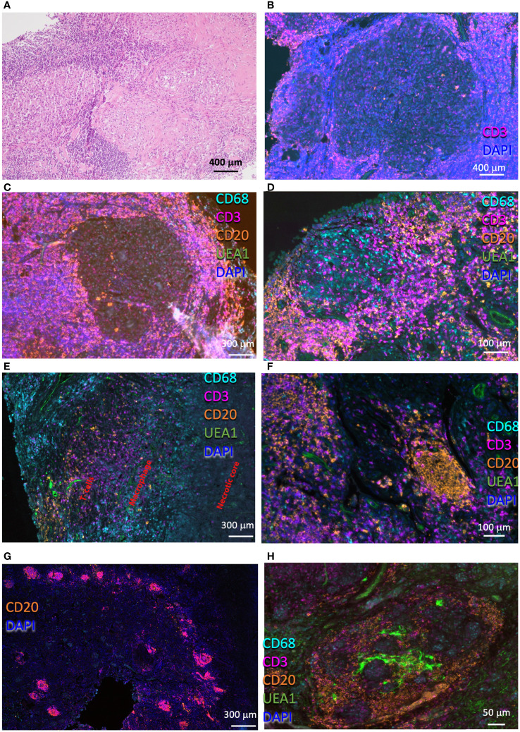
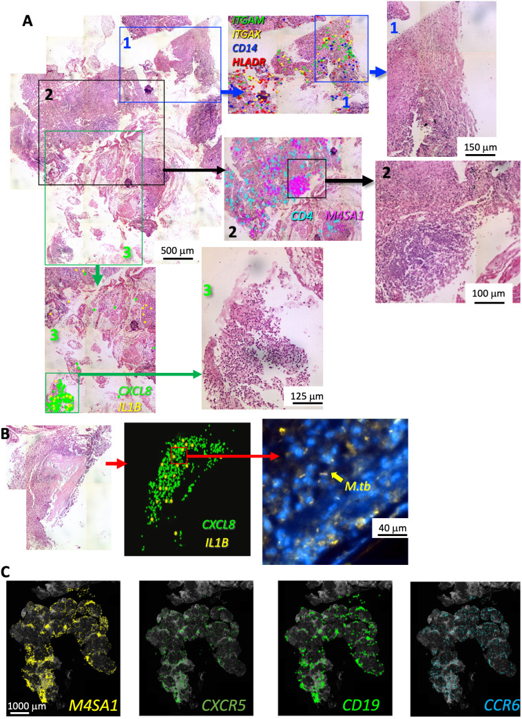
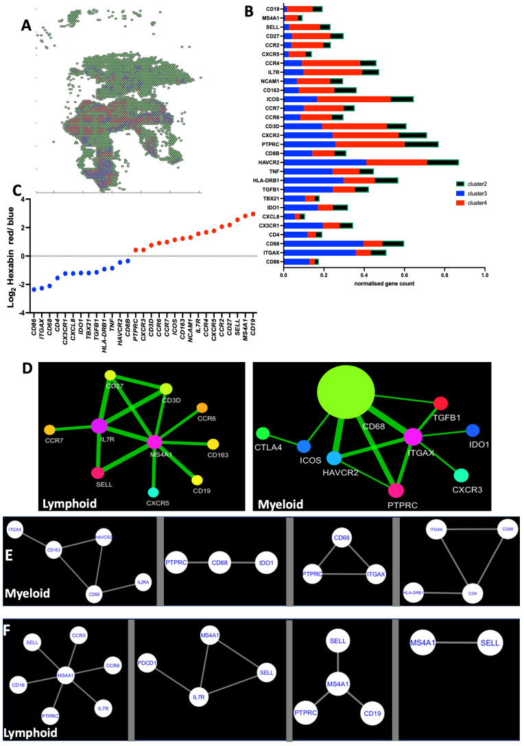
Tuberculosis (TB) and sarcoidosis are both granulomatous diseases. Here, we compared the immunological microenvironments of granulomas from TB and sarcoidosis patients using sequencing (ISS) transcriptomic analysis and multiplexed immunolabeling of tissue sections. TB lesions consisted of large necrotic and cellular granulomas, whereas "multifocal" granulomas with macrophages or epitheloid cell core and a T-cell rim were observed in sarcoidosis samples. The necrotic core in TB lesions was surrounded by macrophages and encircled by a dense T-cell layer. Within the T-cell layer, compact B-cell aggregates were observed in most TB samples. These B-cell clusters were vascularized and could contain defined B-/T-cell and macrophage-rich areas. The ISS of 40-60 immune transcripts revealed the enriched expression of transcripts involved in homing or migration to lymph nodes, which formed networks at single-cell distances in lymphoid areas of the TB lesions. Instead, myeloid-annotated regions were enriched in , , , , and mRNA. and mRNA were observed in granulocytic areas in which was also detected. In line with ISS data indicating tertiary lymphoid structures, immune labeling of TB sections expressed markers of high endothelial venules, follicular dendritic cells, follicular helper T cells, and lymph-node homing receptors on T cells. Neither ISS nor immunolabeling showed evidence of tertiary lymphoid aggregates in sarcoidosis samples. Together, our finding suggests that despite their heterogeneity, the formation of tertiary immune structures is a common feature in granulomas from TB patients.
肺结核(TB)和结节病都是肉芽肿性疾病。在这里,我们使用测序(ISS)转录组分析和组织切片的多重免疫标记比较了来自 TB 和结节病患者的肉芽肿的免疫微环境。TB 病变由大的坏死和细胞性肉芽肿组成,而结节病样本中观察到“多灶性”肉芽肿,其具有巨噬细胞或上皮样细胞核心和 T 细胞边缘。TB 病变中的坏死核心被巨噬细胞包围,并被致密的 T 细胞层包围。在 T 细胞层内,大多数 TB 样本中观察到密集的 B 细胞聚集。这些 B 细胞簇是血管化的,可以包含定义明确的 B/T 细胞和巨噬细胞丰富区。40-60 个免疫转录本的 ISS 显示了参与归巢或迁移到淋巴结的转录本的丰富表达,这些转录本在 TB 病变的淋巴区域以单细胞距离形成网络。相反,髓系注释区域富含 、 、 、 和 mRNA。在粒细胞区域中观察到 和 mRNA,并且也检测到 。与表明三级淋巴结构的 ISS 数据一致,TB 切片的免疫标记表达高内皮静脉、滤泡树突状细胞、滤泡辅助 T 细胞和 T 细胞上的淋巴结归巢受体的标志物。ISS 或免疫标记均未显示结节病样本中存在三级淋巴聚集的证据。总之,我们的发现表明,尽管存在异质性,但三级免疫结构的形成是 TB 患者肉芽肿的共同特征。