Harrell R M, Lyles K W, Harrelson J M, Friedman N E, Drezner M K
J Clin Invest. 1985 Jun;75(6):1858-68. doi: 10.1172/JCI111900.
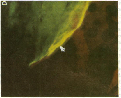
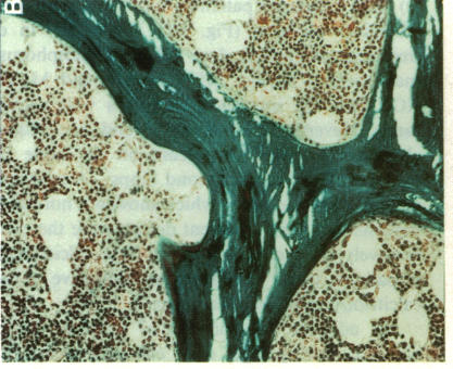
Although conventional therapy (pharmacologic doses of vitamin D and phosphorus supplementation) is usually successful in healing the rachitic bone lesion in patients with X-linked hypophosphatemic rickets, it does not heal the coexistent osteomalacia. Because serum 1,25-dihydroxyvitamin D levels are inappropriately low in these patients and high calcitriol concentrations may be required to heal the osteomalacia, we chose to treat five affected subjects with high doses of calcitriol (68.2 +/- 10.0 ng/kg total body weight/d) and supplemental phosphorus (1-2 g/d) performing metabolic studies and bone biopsies before and after 5-8 mo of this therapy in each individual. Of these five patients, three (aged 13, 13, and 19 yr) were receiving conventional treatment at the inception of the study and therefore showed base-line serum phosphorus concentrations within the normal range. The remaining two untreated patients (aged 2 and 37 yr) displayed characteristic hypophosphatemia before calcitriol therapy. All five patients demonstrated serum calcitriol levels in the low normal range (22.5 +/- 3.2 pg/ml), impaired renal phosphorus conservation (tubular maximum for the reabsorption of phosphate per deciliter of glomerular filtrate, 2.13 +/- 0.20 mg/dl), and osteomalacia on bone biopsy (relative osteoid volume, 14.4 +/- 1.7%; mean osteoid seam width, 27.7 +/- 3.7 micron; mineral apposition rate, 0.46 +/- 0.12 micron/d). On high doses of calcitriol, serum 1,25-dihydroxyvitamin D levels rose into the supraphysiologic range (74.1 +/- 3.8 pg/ml) with an associated increment in the serum phosphorus concentration (2.82 +/- 0.19 to 3.78 +/- 0.32 mg/dl) and improvement of the renal tubular maximum for phosphate reabsorption (3.17 +/- 0.22 mg/dl). The serum calcium rose in each patient while the immunoactive parathyroid hormone concentration measured by three different assays remained within the normal range. Most importantly, repeat bone biopsies showed that high doses of calcitriol and phosphorus supplements had reversed the mineralization defect in all patients (mineral apposition rate, 0.88 +/- 0.04 micron/d) and consequently reduced parameters of bone osteoid content to normal (relative osteoid volume, 4.1 +/- 0.7%; mean osteoid seam width, 11.0 +/- 1.0 micron). Complications (hypercalcemia and hypercalciuria) ensued in four of these five patients within 1-17 mo of documented bone healing, necessitating reduction of calcitriol doses to a mean of 1.6 +/- 0.2 micrograms/d (28 +/- 4 ng/kg ideal body weight per day). At follow-up bone biopsy, these four subjects continued to manifest normal bone mineralization dynamics (mineral apposition rate, 0.88 +/-0.10 micrometer/d) on reduced doses of 1.25-dihydroxyvitamin D with phosphorus supplements (2 g/d) for a mean of 21.3 +/- 1.3 mo after bone healing was first documented. Static histomorphometric parameters also remained normal (relative osteoid volume, 1.5 +/- 0.4%; mean osteoid seam width, 13.5 +/- 0.8 micrometer). These data indicate that administration of supraphysiologic amounts of calcitriol, in conjunction with oral phosphorus, results in complete healing of vitamin D resistant osteomalacia in patients with X-linked hypophosphatemic rickets. Although complications predictably require calcitriol dose reductions once healing is achieved, continued bone healing can be maintained for up to 1 yr with lower doses of 1,25-dihydroxyvitamin D and continued phosphorus supplementation.
虽然传统疗法(药理剂量的维生素D和补充磷)通常能成功治愈X连锁低磷性佝偻病患者的佝偻病性骨病变,但无法治愈并存的骨软化症。由于这些患者的血清1,25-二羟维生素D水平异常低,可能需要高浓度的骨化三醇来治愈骨软化症,我们选择用高剂量骨化三醇(68.2±10.0 ng/kg体重/天)和补充磷(1 - 2 g/天)治疗5名受影响的受试者,并在每个个体接受该治疗5 - 8个月前后进行代谢研究和骨活检。这5名患者中,3名(年龄分别为13岁、13岁和19岁)在研究开始时接受传统治疗,因此基线血清磷浓度在正常范围内。其余2名未治疗的患者(年龄分别为2岁和37岁)在骨化三醇治疗前表现出典型的低磷血症。所有5名患者的血清骨化三醇水平均处于低正常范围(22.5±3.2 pg/ml),肾磷保留功能受损(每分升肾小球滤过液中磷的肾小管最大重吸收率,2.13±0.20 mg/dl),骨活检显示存在骨软化症(相对类骨质体积,14.4±1.7%;平均类骨质缝宽度,27.7±3.7微米;矿物质沉积率,0.46±0.12微米/天)。给予高剂量骨化三醇后,血清1,25-二羟维生素D水平升至超生理范围(74.1±3.8 pg/ml),血清磷浓度随之升高(从2.82±0.19升至3.78±0.32 mg/dl),肾小管最大磷重吸收率得到改善(3.17±0.22 mg/dl)。每位患者的血清钙均升高,而通过三种不同检测方法测得的免疫活性甲状旁腺激素浓度仍在正常范围内。最重要的是,重复骨活检显示,高剂量骨化三醇和补充磷使所有患者的矿化缺陷得到逆转(矿物质沉积率,0.88±0.04微米/天),从而使骨类骨质含量参数恢复正常(相对类骨质体积,4.1±0.7%;平均类骨质缝宽度,11.0±1.0微米)。这5名患者中有4名在记录到骨愈合后的1 - 17个月内出现并发症(高钙血症和高钙尿症),因此需要将骨化三醇剂量减至平均1.6±0.2微克/天(28±4 ng/kg理想体重/天)。在随访骨活检时,这4名受试者在首次记录到骨愈合后,平均21.3±1.3个月内,在减少剂量的1,25-二羟维生素D和补充磷(2 g/天)治疗下,继续表现出正常的骨矿化动态(矿物质沉积率,0.88±0.10微米/天)。静态组织形态计量学参数也保持正常(相对类骨质体积,1.5±0.4%;平均类骨质缝宽度,13.5±0.8微米)。这些数据表明,给予超生理量的骨化三醇并联合口服磷,可使X连锁低磷性佝偻病患者的维生素D抵抗性骨软化症完全愈合。虽然一旦愈合,并发症可预见地需要减少骨化三醇剂量,但较低剂量的1,25-二羟维生素D和持续补充磷可使骨愈合持续维持长达1年。