Ahn WonMo, Burnett Faith N, Pandey Ajay, Ghoshal Pushpankur, Singla Bhupesh, Simon Abigayle B, Derella Cassandra C, A Addo Stephen, Harris Ryan A, Lucas Rudolf, Csányi Gábor
Vascular Biology Center, Medical College of Georgia, Augusta University, Augusta, GA 30912, USA.
Georgia Prevention Institute, Medical College of Georgia, Augusta University, Augusta, GA 30912, USA.
Antioxidants (Basel). 2024 Jan 30;13(2):175. doi: 10.3390/antiox13020175.
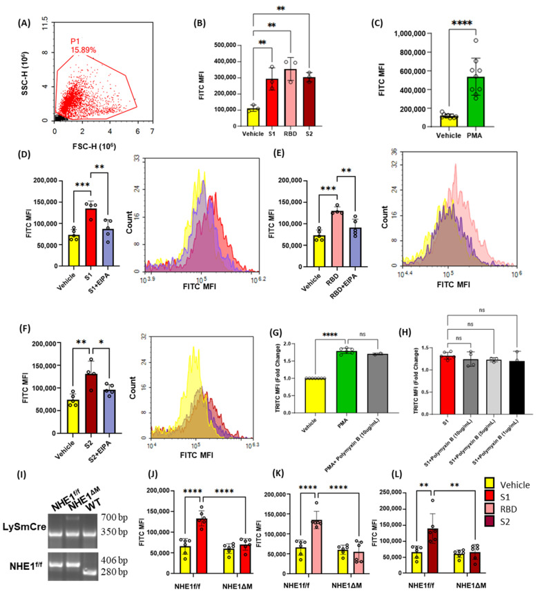
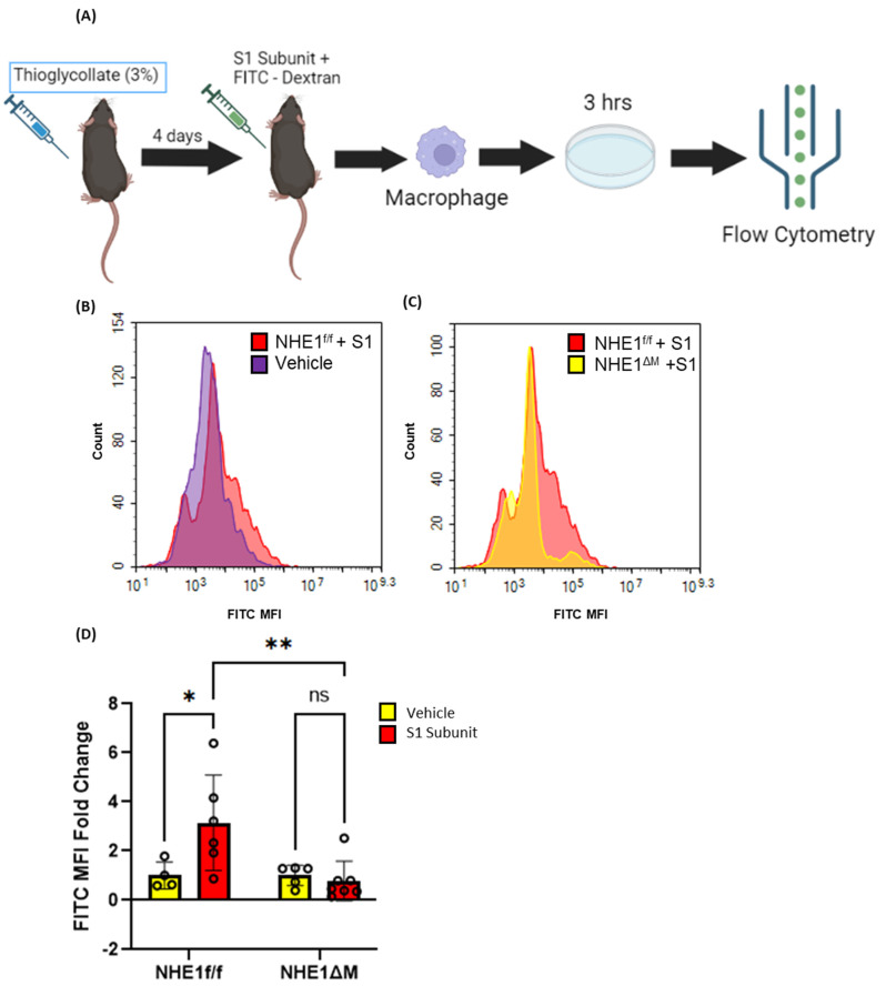
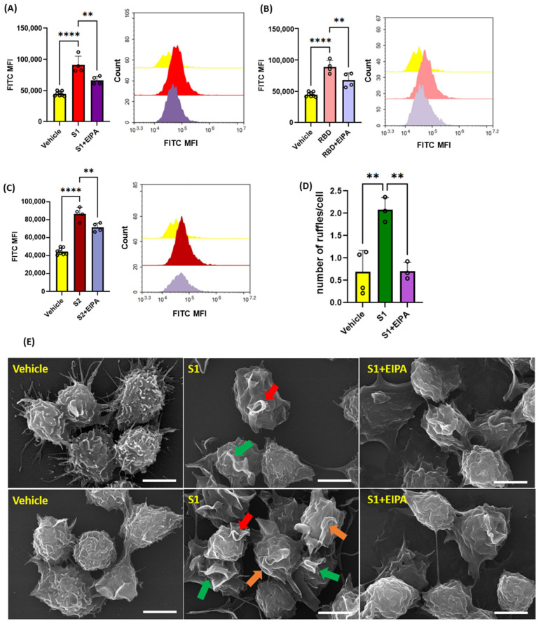
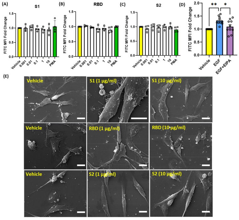
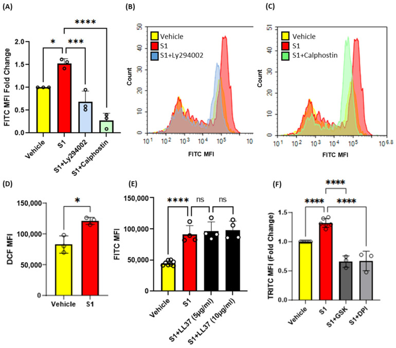
Coronavirus disease 2019 (COVID-19) is an infectious disease caused by severe acute respiratory syndrome coronavirus 2 (SARS-CoV-2). While recent studies have demonstrated that SARS-CoV-2 may enter kidney and colon epithelial cells by inducing receptor-independent macropinocytosis, it remains unknown whether this process also occurs in cell types directly relevant to SARS-CoV-2-associated lung pneumonia, such as alveolar epithelial cells and macrophages. The goal of our study was to investigate the ability of SARS-CoV-2 spike protein subunits to stimulate macropinocytosis in human alveolar epithelial cells and primary human and murine macrophages. Flow cytometry analysis of fluid-phase marker internalization demonstrated that SARS-CoV-2 spike protein subunits S1, the receptor-binding domain (RBD) of S1, and S2 stimulate macropinocytosis in both human and murine macrophages in an angiotensin-converting enzyme 2 (ACE2)-independent manner. Pharmacological and genetic inhibition of macropinocytosis substantially decreased spike-protein-induced fluid-phase marker internalization in macrophages both in vitro and in vivo. High-resolution scanning electron microscopy (SEM) imaging confirmed that spike protein subunits promote the formation of membrane ruffles on the dorsal surface of macrophages. Mechanistic studies demonstrated that SARS-CoV-2 spike protein stimulated macropinocytosis via NADPH oxidase 2 (Nox2)-derived reactive oxygen species (ROS) generation. In addition, inhibition of protein kinase C (PKC) and phosphoinositide 3-kinase (PI3K) in macrophages blocked SARS-CoV-2 spike-protein-induced macropinocytosis. To our knowledge, these results demonstrate for the first time that SARS-CoV-2 spike protein subunits stimulate macropinocytosis in macrophages. These results may contribute to a better understanding of SARS-CoV-2 infection and COVID-19 pathogenesis.
2019冠状病毒病(COVID-19)是由严重急性呼吸综合征冠状病毒2(SARS-CoV-2)引起的一种传染病。虽然最近的研究表明,SARS-CoV-2可能通过诱导非受体依赖性巨胞饮作用进入肾脏和结肠上皮细胞,但尚不清楚这一过程是否也发生在与SARS-CoV-2相关的肺炎直接相关的细胞类型中,如肺泡上皮细胞和巨噬细胞。我们研究的目的是调查SARS-CoV-2刺突蛋白亚基刺激人肺泡上皮细胞以及原代人巨噬细胞和小鼠巨噬细胞发生巨胞饮作用的能力。对液相标记内化的流式细胞术分析表明,SARS-CoV-2刺突蛋白亚基S1、S1的受体结合域(RBD)和S2以不依赖血管紧张素转换酶2(ACE2)的方式刺激人和小鼠巨噬细胞发生巨胞饮作用。巨胞饮作用的药理学和遗传学抑制在体外和体内均显著降低了刺突蛋白诱导的巨噬细胞液相标记内化。高分辨率扫描电子显微镜(SEM)成像证实,刺突蛋白亚基促进巨噬细胞背表面膜皱褶的形成。机制研究表明,SARS-CoV-2刺突蛋白通过烟酰胺腺嘌呤二核苷酸磷酸(NADPH)氧化酶2(Nox2)产生的活性氧(ROS)刺激巨胞饮作用。此外,巨噬细胞中蛋白激酶C(PKC)和磷脂酰肌醇3激酶(PI3K)的抑制阻断了SARS-CoV-2刺突蛋白诱导的巨胞饮作用。据我们所知,这些结果首次证明SARS-CoV-2刺突蛋白亚基刺激巨噬细胞发生巨胞饮作用。这些结果可能有助于更好地理解SARS-CoV-2感染和COVID-19的发病机制。