Department of Clinical Sciences, Obstetric, Gynecological and Prenatal Ultrasound Research, Lund University, Malmö, Sweden.
Department of Obstetrics and Gynecology, Skåne University Hospital, Malmö, Sweden.
Acta Obstet Gynecol Scand. 2024 Jun;103(6):1142-1152. doi: 10.1111/aogs.14812. Epub 2024 Feb 27.
Studies that use standardized ultrasonographic criteria to diagnose adenomyosis in subfertile women are needed. These would improve the understanding of the disease burden and enable further studies on its impact on fertility and assisted reproductive treatment (ART) outcome. The aim of this study was to determine the prevalence of different features of adenomyosis in women scheduled for their first ART, diagnosed at two (2D) and three-dimensional (3D) transvaginal ultrasonography (TVUS) using the revised Morphological Uterus Sonographic Assessment (MUSA) group definitions.
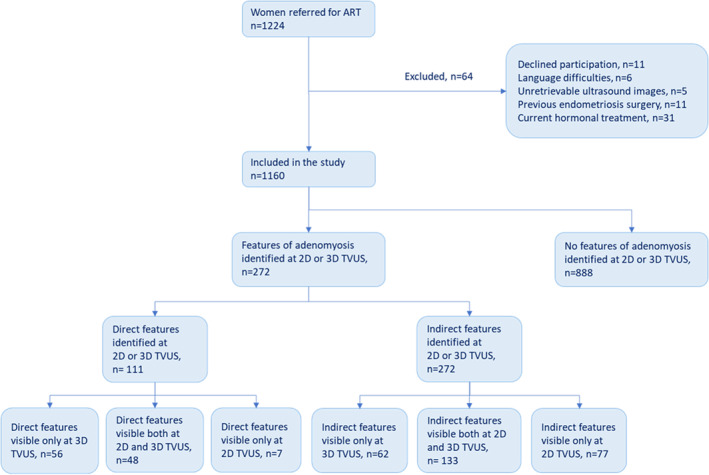
This was a prospective, observational cross-sectional study of subfertile women aged 25 to ≤39 years, that were referred to a university hospital for their first ART between December 2018 and May 2021. Of 1224 eligible women, 1160 women fulfilled the inclusion criteria and consented to participate in the study. All women underwent a systematic 2D and 3D TVUS examination. The primary outcome was the presence of direct and indirect features of adenomyosis, as proposed by the MUSA group. Secondary outcomes were to describe the ultrasonographic characteristics of the different features, as well as any difference in the diagnostics at 2D or 3D TVUS and any association with clinical characteristics such as endometriosis.
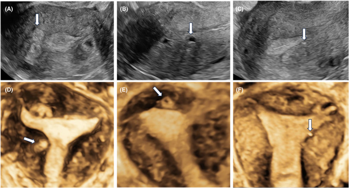
At least one direct or indirect feature of adenomyosis was observed in 272 (23.4%, 95% confidence interval [CI] 21.0-25.9) women. Direct features that are pathognomonic for the disease were observed in 111 (9.6%, 95% CI, 7.9-11.3) women. Direct features were visible only at 3D TVUS in 56 (4.8%, 95% CI 3.6-6.1) women, that is, 56/111 (50.5%) of women with at least one direct adenomyosis feature. Direct features were more common in women with endometriosis (OR 2.8, 95% CI 1.8-4.3).
We found than one in 10 women scheduled for ART had direct features of adenomyosis at ultrasound examination. The present study suggests that the use of 3D TVUS is an important complement to 2D in the diagnostics of adenomyosis. Our results may further improve the counseling of women scheduled for ART and enables future studies on the impact of different features of adenomyosis on subfertility, ART results and obstetric outcomes.
需要进行研究,以标准化的超声标准诊断不孕妇女的子宫腺肌病。这将有助于更好地了解疾病负担,并能够进一步研究其对生育能力和辅助生殖治疗(ART)结果的影响。本研究旨在通过使用修订后的形态子宫超声评估(MUSA)组定义,在接受首次 ART 的女性中,通过二维(2D)和三维(3D)经阴道超声(TVUS)检查,确定不同类型的子宫腺肌病的患病率。
这是一项前瞻性、观察性的横断性研究,纳入了年龄在 25 至 39 岁之间、2018 年 12 月至 2021 年 5 月期间因首次 ART 而被转诊至大学医院的不孕妇女。在 1224 名符合条件的女性中,有 1160 名女性符合纳入标准并同意参与研究。所有女性均接受了系统的 2D 和 3D TVUS 检查。主要结局是观察到 MUSA 组提出的直接和间接的子宫腺肌病特征。次要结局是描述不同特征的超声特征,以及 2D 或 3D TVUS 检查的诊断差异,以及与子宫内膜异位症等临床特征的任何关联。
在 272 名(23.4%,95%置信区间 [CI] 21.0-25.9)女性中观察到至少一种直接或间接的子宫腺肌病特征。在 111 名(9.6%,95%CI 7.9-11.3)女性中观察到与疾病相关的直接特征。在 56 名(4.8%,95%CI 3.6-6.1)女性中仅在 3D TVUS 上可见直接特征,即 111 名有至少一种直接子宫腺肌病特征的女性中有 56 名(50.5%)。在患有子宫内膜异位症的女性中,直接特征更为常见(OR 2.8,95%CI 1.8-4.3)。
我们发现,在接受 ART 治疗的女性中,有十分之一以上的女性在超声检查中存在直接的子宫腺肌病特征。本研究表明,在诊断子宫腺肌病方面,3D TVUS 是 2D 的重要补充。我们的研究结果可能会进一步改善对接受 ART 治疗的女性的咨询,并能够进一步研究不同类型的子宫腺肌病对不孕、ART 结果和产科结局的影响。