Celardo Gaetano, Scaffei Elena, Buchignani Bianca, Donatelli Graziella, Costagli Mauro, Cristofani Paola, Canapicchi Raffaello, Pasquariello Rosa, Tosetti Michela, Battini Roberta, Biagi Laura
Department of Developmental Neuroscience, IRCCS Stella Maris Foundation, Pisa, Italy.
Department of Clinical and Experimental Medicine, University of Pisa, Pisa, Italy.
Front Neurol. 2024 Feb 14;15:1362704. doi: 10.3389/fneur.2024.1362704. eCollection 2024.
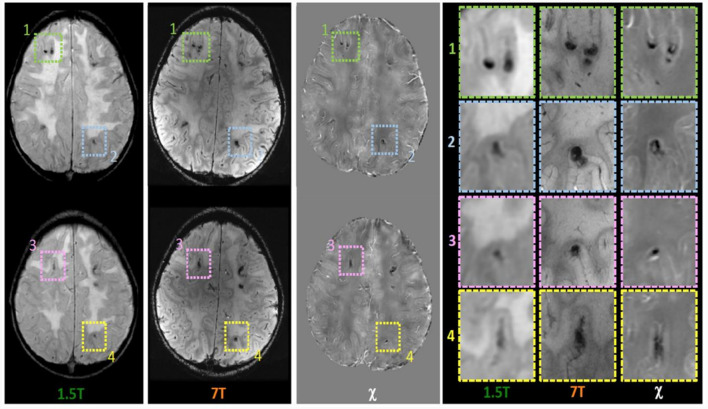
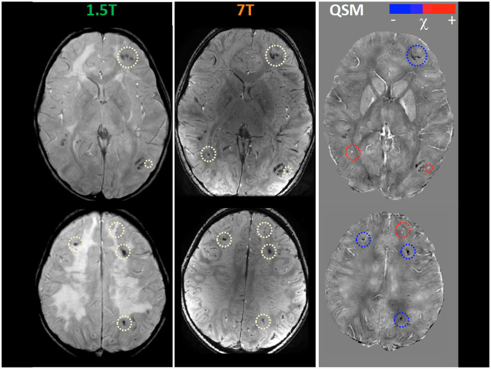
Chemotherapy and radiotherapy are widely used in the treatment of central nervous system tumors and acute lymphocytic leukemia even in the pediatric population. However, such treatments run the risk of a broad spectrum of cognitive and neurological deficits. Even though the correlation with cognitive decline is still not clear, neuroradiological defects linked to white matter injury and vasculopathies may be identified. Thanks to the use of 7T MRI it is possible to better define the vascular pattern of the brain lesions with the added advantage of identifying their characteristics and anatomical localization, which, however, are not evident with a conventional brain scan. Moreover, the use of Quantitative Susceptibility Mapping (QSM) makes it possible to discriminate between calcium deposits on vessels (chemo-radiation-induced) and hemoglobin deposition in radio-induced cavernomas, speculating, as a result, about the pathophysiology of iatrogenic brain damage. We describe the case of a 9 year-old boy with a T-type acute lymphoid leukemia who had previously been treated with polychemotherapy and high-dose RT. To better define the child's neuroradiological pattern, 7T MRI and QSM were performed in addition to conventional imaging examinations. Our case report suggests the potential usefulness of a QSM study to distinguish radio-induced vascular malformations from mineralizing microangiopathy.
化疗和放疗广泛应用于中枢神经系统肿瘤和急性淋巴细胞白血病的治疗,甚至在儿科患者中也是如此。然而,这类治疗存在引发广泛认知和神经功能缺损的风险。尽管与认知衰退的关联仍不明确,但与白质损伤和血管病变相关的神经放射学缺陷可能会被识别出来。借助7T磁共振成像(MRI),能够更好地界定脑部病变的血管形态,还有额外的优势,即识别其特征和解剖定位,而这些在传统脑部扫描中并不明显。此外,使用定量磁化率图谱(QSM)可以区分血管上的钙沉积(化疗放疗诱导)和放射性海绵状血管瘤中的血红蛋白沉积,从而推测医源性脑损伤的病理生理学。我们描述了一名9岁患T型急性淋巴细胞白血病男孩的病例,该男孩此前接受过联合化疗和高剂量放疗。为了更好地界定该患儿的神经放射学特征,除了进行传统成像检查外,还进行了7T MRI和QSM检查。我们的病例报告表明,QSM研究在区分放射性血管畸形和矿化性微血管病方面可能具有潜在用途。