Ramalho Rodrigues Rodrigo, Neto Diógenes Firmino do Nascimento, Andrade Fernandes João Vítor, Barreto Letícia de Oliveira, Barros Maciel do Amaral Victor, Karoline de Araújo Deca Débora, Freire de Albuquerque Figueiredo Vera Louise, Dantas de Lucena Jalles, Bezerra da Silva Ivson, Sales Thales Henrique de Araújo, Oliveira André de Sá Braga
Federal University of Paraiba, João Pessoa, Brazil.
Centro Universitário Santa Maria, Cajazeiras, Brazil.
Anat Cell Biol. 2024 Jun 30;57(2):213-220. doi: 10.5115/acb.23.218. Epub 2024 Mar 7.
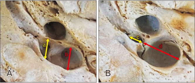
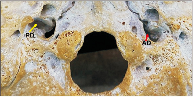
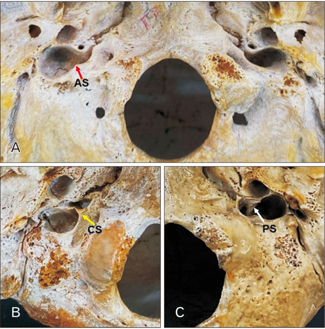
The jugular foramen (JF) is located between the temporal and occipital bones. The JF is a primary pathway for venous outflow from the skull and passage of nerves. Variations are common in this region and may have clinical and surgical implications. To analyze the sexual dimorphism and JF morphology in skulls from Northeastern Brazil. 128 human skulls from the Anatomy Laboratory of the Federal University of Paraíba, 64 male and 64 female, were selected and the JFs analyzed for bone septation and the presence of a dome. Data analysis considered <0.05 as significant. On at least one side, complete septation was observed in 26 skulls (20.3%), incomplete septation in 93 skulls (72.6%) and 61 skulls (47.6%) did not present septation. In 114 skulls (89%), 47.6% female and 41.4% male, have a unilateral presence of the dome and 71 (55.4%) have it bilaterally. Posterolateral compartment diameters and JF area had higher values on the right side in the total sample and separated by sex (<0.05). Most morphometric variables of the anteromedial compartment were higher in male than in female (<0.05), fact that was not observed in the posterolateral compartment (>0.05). This study showed a higher prevalence of complete septation in males compared to females. Morphometric analysis presented a peculiar morphology of the JF in this study. These results suggests that the surgical approach to diseases that affect the JF may be peculiar to the studied population, confirming the importance of morphological analysis of the skull base.
颈静脉孔(JF)位于颞骨和枕骨之间。颈静脉孔是颅骨静脉流出和神经通过的主要通道。该区域变异常见,可能具有临床和手术意义。为分析巴西东北部颅骨的性别二态性和颈静脉孔形态。从帕拉伊巴联邦大学解剖实验室选取了128具人类颅骨,其中男性64具,女性64具,并对颈静脉孔进行骨分隔和穹窿存在情况分析。数据分析以<0.05为显著水平。至少在一侧,26具颅骨(20.3%)观察到完全分隔,93具颅骨(72.6%)为不完全分隔,另有61具颅骨(47.6%)未出现分隔。114具颅骨(89%),其中女性占47.6%,男性占41.4%,存在单侧穹窿,71具颅骨(55.4%)双侧存在穹窿。总样本中右侧后外侧隔室直径和颈静脉孔面积值更高,且按性别区分有差异(<0.05)。前内侧隔室的大多数形态测量变量男性高于女性(<0.05),而后外侧隔室未观察到这种情况(>0.05)。本研究表明男性完全分隔的患病率高于女性。形态测量分析显示本研究中颈静脉孔具有独特形态。这些结果表明,针对影响颈静脉孔疾病的手术入路可能因研究人群而异,证实了颅底形态分析的重要性。