Cortez Thiago Vinícius, Cerqueira Nathália Mancioppi, Gallas Julia Adornes, Oliveira Wanderley Pereira, Corona Silmara Aparecida Milori, Souza-Gabriel Aline Evangelista
Department of Restorative Dentistry, School of Dentistry of Ribeirão Preto, University of São Paulo, Ribeirão Preto, SP, Brazil.
Department of Pharmaceutical Sciences, School of Pharmaceutical Sciences of Ribeirão Preto, University of São Paulo, Ribeirão Preto, SP, Brazil.
Restor Dent Endod. 2024 Jan 26;49(1):e9. doi: 10.5395/rde.2024.49.e9. eCollection 2024 Feb.
This study aimed to evaluate the effect of pomegranate solution () on eroded dentin through antioxidant action, shear bond strength (SBS) and interface morphology.
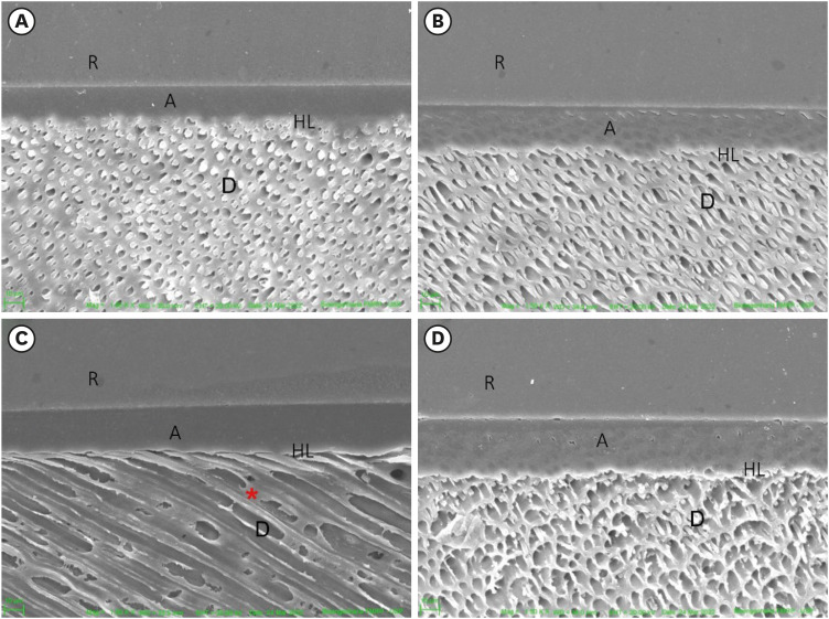
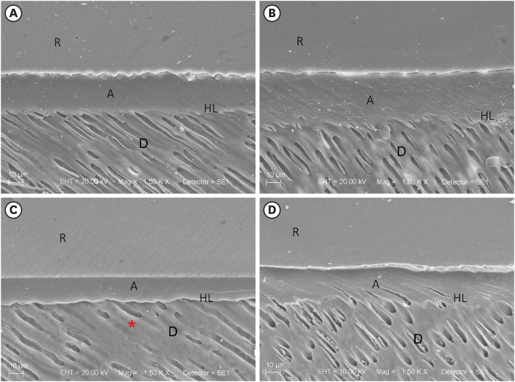
The 10% pomegranate peel extract was prepared by the lyophilization method. Punicalagin polyphenol was confirmed by high-performance liquid chromatography. Antioxidant activity was evaluated by capturing the 2,2-diphenyl-1-picrylhydrazyl (DPPH) radical. For the SBS, 48 dentin fragments were divided into sound or eroded, and subdivided according to the pretreatment ( = 12): water or . The surfaces were restored with self-etch adhesive and a bulk-fill resin (Ecosite; DMG). The SBS was done immediately (24 hours) and after thermal cycling + water storage (12 months). For scanning electron microscopy, 48 dentin fragments (24 sound and 24 eroded) received the same treatments as for SBS ( = 6), and they were analyzed after 24 hours and 12 months.
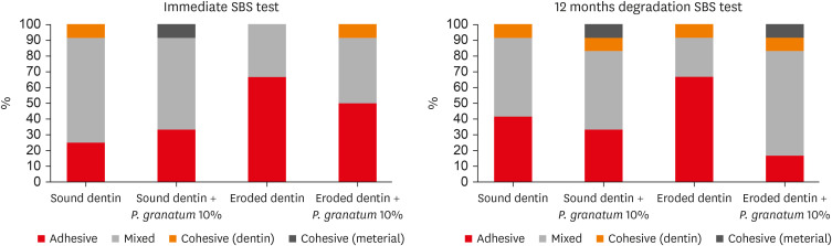
The had antioxidant action similar ( = 0.246) to the phenolic standard antioxidants. After 24 hours, eroded dentin had lower SBS than sound dentin ( < 0.001), regardless of the pretreatment. After 12 months, maintained the SBS of sound dentin (13.46 ± 3.42 MPa) and eroded dentin (10.96 ± 1.90 MPa) statistically similar. The lowest values were found on eroded dentin treated with water (5.75 ± 1.65 MPa) ( < 0.001). on eroded dentin caused peritubular demineralization and hybrid layer with resin tags.
The pomegranate extract had antioxidant action and preserved the adhesive interface of the eroded dentin.
本研究旨在通过抗氧化作用、剪切粘结强度(SBS)和界面形态评估石榴溶液对侵蚀性牙本质的影响。
采用冻干法制备10%石榴皮提取物。通过高效液相色谱法确认石榴皮素多酚。通过捕获2,2-二苯基-1-苦基肼(DPPH)自由基评估抗氧化活性。对于SBS,将48个牙本质碎片分为完好或侵蚀性的,并根据预处理方式(n = 12)进一步细分:水或石榴溶液。表面用自酸蚀粘结剂和大容量填充树脂(Ecosite;DMG)修复。SBS在即刻(24小时)以及热循环+水储存(12个月)后进行检测。对于扫描电子显微镜检查,48个牙本质碎片(24个完好的和24个侵蚀性的)接受与SBS相同的处理(n = 6),并在24小时和12个月后进行分析。
石榴溶液具有与酚类标准抗氧化剂相似(p = 0.246)的抗氧化作用。24小时后,无论预处理方式如何,侵蚀性牙本质的SBS均低于完好牙本质(p < 0.001)。12个月后,石榴溶液使完好牙本质(13.46 ± 3.42 MPa)和侵蚀性牙本质(10.96 ± 1.90 MPa)的SBS在统计学上相似。用水处理的侵蚀性牙本质的SBS值最低(5.75 ± 1.65 MPa)(p < 0.001)。石榴溶液处理侵蚀性牙本质导致管周脱矿和带有树脂突的混合层。
石榴提取物具有抗氧化作用,并保留了侵蚀性牙本质的粘结界面。