Dental Research Center, Dentistry Research Institute, Tehran University of Medical Sciences, Tehran, Iran.
Department of Microbiology, School of Medicine, Tehran University of Medical Sciences, Tehran, Iran.
BMC Oral Health. 2024 Mar 7;24(1):311. doi: 10.1186/s12903-024-04062-7.
This study was conducted to investigate the efficiency of periodontal ligament (PDL) stem cell-derived exosome-loaded Emodin (Emo@PDL-Exo) in antimicrobial photodynamic therapy (aPDT) on Streptococcus mutans and Lactobacillus acidophilus as the cariogenic bacteria.
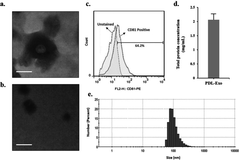
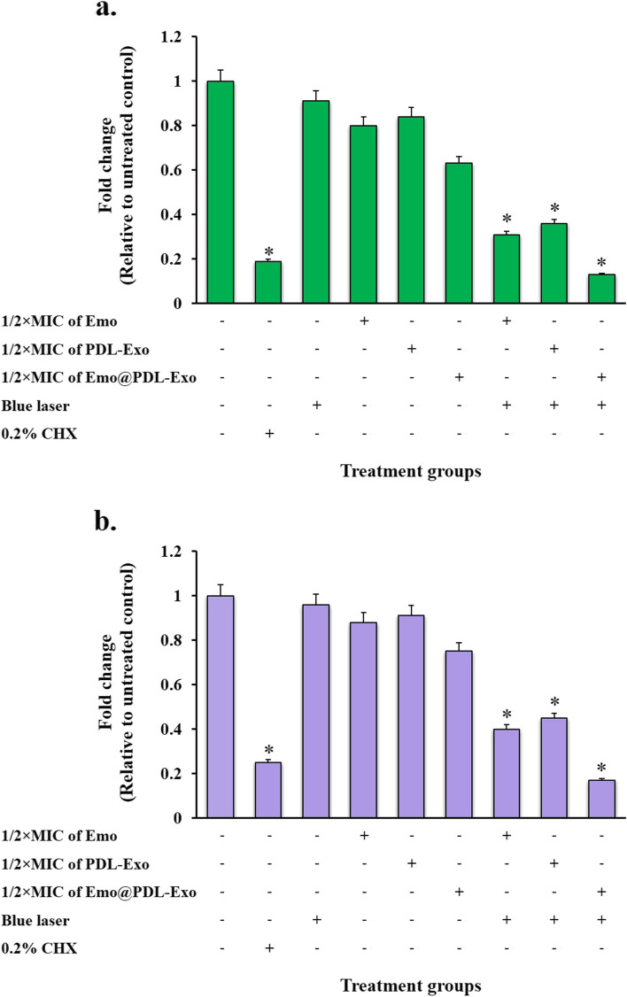
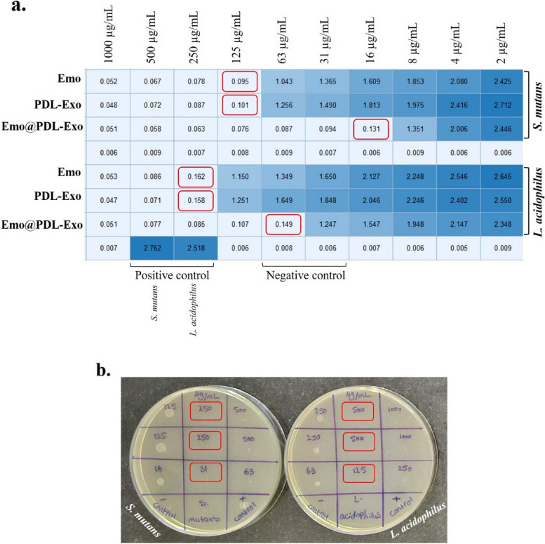
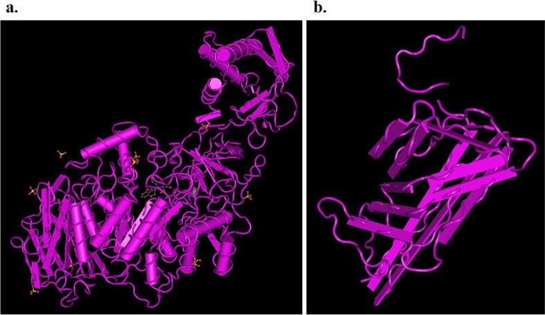
After isolating and characterizing PDL-Exo, the study proceeded to prepare and verify the presence of Emo@PDL-Exo. The antimicrobial effect, anti-biofilm activity, and anti-metabolic potency of Emo, PDL-Exo, and Emo@PDL-Exo were then evaluated with and without irradiation of blue laser at a wavelength of 405 ± 10 nm with an output intensity of 150 mW/cm for a duration of 60 s. In addition, the study assessed the binding affinity of Emodin with GtfB and SlpA proteins using in silico molecular docking. Eventually, the study examined the generation of endogenous reactive oxygen species (ROS) and changes in the gene expression levels of gelE and sprE.
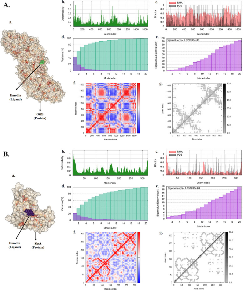
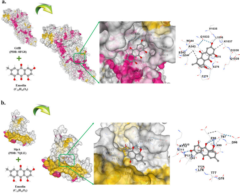
The study found that using Emo@PDL-Exo-mediated aPDT resulted in a significant decrease in L. acidophilus and S. mutans by 4.90 ± 0.36 and 5.07 log CFU/mL, respectively (P < 0.05). The study found that using Emo@PDL-Exo for aPDT significantly reduced L. acidophilus and S. mutans biofilms by 44.7% and 50.4%, respectively, compared to untreated biofilms in the control group (P < 0.05). Additionally, the metabolic activity of L. acidophilus and S. mutans decreased by 58.3% and 71.2%, respectively (P < 0.05). The molecular docking analysis showed strong binding affinities of Emodin with SlpA and GtfB proteins, with docking scores of -7.4 and -8.2 kcal/mol, respectively. The study also found that the aPDT using Emo@PDL-Exo group resulted in the most significant reduction in gene expression of slpA and gtfB, with a decrease of 4.2- and 5.6-folds, respectively, compared to the control group (P < 0.05), likely due to the increased generation of endogenous ROS.
The study showed that aPDT using Emo@PDL-Exo can effectively reduce the cell viability, biofilm activity, and metabolic potency of S. mutans and L. acidophilus. aPDT also significantly reduced the expression levels of gtfB and slpA mRNA due to the increased endogenous ROS generation. The findings suggest that Emo@PDL-Exo-mediated aPDT could be a promising antimicrobial approach against cariogenic microorganisms.
本研究旨在探讨牙周膜干细胞衍生的外泌体负载大黄素(Emo@PDL-Exo)在变形链球菌和嗜酸乳杆菌作为致龋菌的抗菌光动力疗法(aPDT)中的效率。
在分离和表征 PDL-Exo 后,本研究继续制备和验证 Emo@PDL-Exo 的存在。然后,在波长为 405±10nm、输出强度为 150mW/cm 的蓝色激光照射下,评估 Emo、PDL-Exo 和 Emo@PDL-Exo 的抗菌效果、抗生物膜活性和抗代谢活性。此外,本研究使用计算机分子对接评估大黄素与 GtfB 和 SlpA 蛋白的结合亲和力。最终,本研究检测了内源性活性氧(ROS)的产生和 gelE 和 sprE 基因表达水平的变化。
本研究发现,使用 Emo@PDL-Exo 介导的 aPDT 可使嗜酸乳杆菌和变形链球菌分别减少 4.90±0.36 和 5.07 log CFU/mL(P<0.05)。与对照组未处理的生物膜相比,本研究发现使用 Emo@PDL-Exo 进行 aPDT 可使嗜酸乳杆菌和变形链球菌生物膜分别减少 44.7%和 50.4%(P<0.05)。此外,嗜酸乳杆菌和变形链球菌的代谢活性分别降低了 58.3%和 71.2%(P<0.05)。分子对接分析表明,大黄素与 SlpA 和 GtfB 蛋白具有很强的结合亲和力,对接评分分别为-7.4 和-8.2 kcal/mol。本研究还发现,与对照组相比,Emo@PDL-Exo 组的 aPDT 导致 slpA 和 gtfB 基因表达显著降低,分别降低了 4.2-和 5.6 倍(P<0.05),这可能是由于内源性 ROS 的产生增加所致。
本研究表明,Emo@PDL-Exo 介导的 aPDT 可有效降低变形链球菌和嗜酸乳杆菌的细胞活力、生物膜活性和代谢活力。aPDT 还因内源性 ROS 的产生增加而显著降低 gtfB 和 slpA mRNA 的表达水平。研究结果表明,Emo@PDL-Exo 介导的 aPDT 可能是一种有前途的抗致龋微生物的抗菌方法。