Milano Serena, Saponara Ilenia, Gerbino Andrea, Lapi Dominga, Lela Ludovica, Carmosino Monica, Dal Monte Massimo, Bagnoli Paola, Svelto Maria, Procino Giuseppe
Department of Biosciences, Biotechnologies and Environment, University of Bari, Bari, Italy.
Department of Biology, University of Pisa, Pisa, Italy.
Front Physiol. 2024 Feb 22;15:1304375. doi: 10.3389/fphys.2024.1304375. eCollection 2024.
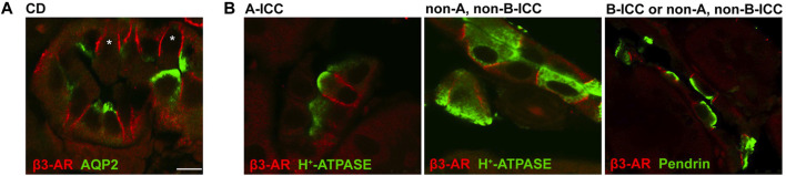
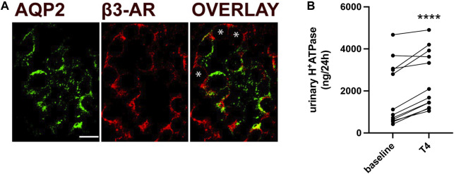
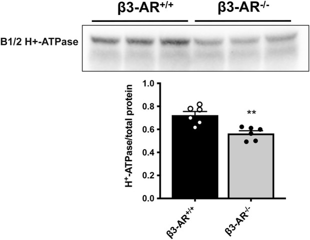
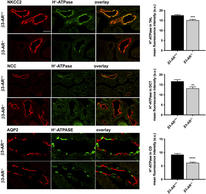
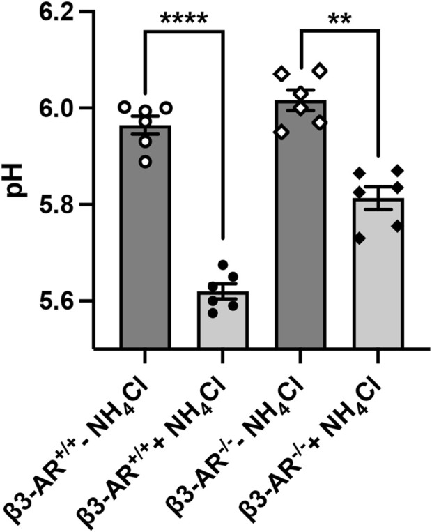
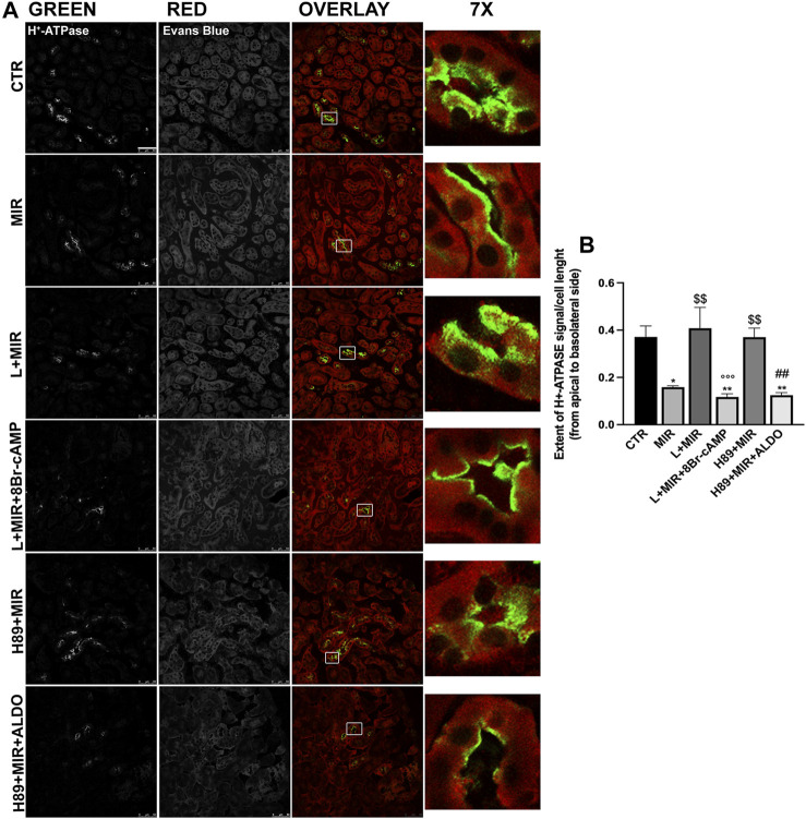
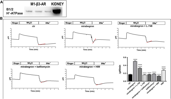
Efferent sympathetic nerve fibers regulate several renal functions activating norepinephrine receptors on tubular epithelial cells. Of the beta-adrenoceptors (β-ARs), we previously demonstrated the renal expression of β3-AR in the thick ascending limb (TAL), the distal convoluted tubule (DCT), and the collecting duct (CD), where it participates in salt and water reabsorption. Here, for the first time, we reported β3-AR expression in the CD intercalated cells (ICCs), where it regulates acid-base homeostasis. Co-localization of β3-AR with either proton pump H-ATPase or Cl/HCO exchanger pendrin revealed β3-AR expression in type A, type B, non-A, and non-B ICCs in the mouse kidney. We aimed to unveil the possible regulatory role of β3-AR in renal acid-base homeostasis, in particular in modulating the expression, subcellular localization, and activity of the renal H-ATPase, a key player in this process. The abundance of H-ATPase was significantly decreased in the kidneys of β3-AR compared with those of β3-AR mice. In particular, H-ATPase reduction was observed not only in the CD but also in the TAL and DCT, which contribute to acid-base transport in the kidney. Interestingly, we found that in , the absence of β3-AR reduced the kidneys' ability to excrete excess proton in the urine during an acid challenge. Using stimulation of mouse kidney slices, we proved that the β3-AR activation promoted H-ATPase apical expression in the epithelial cells of β3-AR-expressing nephron segments, and this was prevented by β3-AR antagonism or PKA inhibition. Moreover, we assessed the effect of β3-AR stimulation on H-ATPase activity by measuring the intracellular pH recovery after an acid load in β3-AR-expressing mouse renal cells. Importantly, β3-AR agonism induced a 2.5-fold increase in H-ATPase activity, and this effect was effectively prevented by β3-AR antagonism or by inhibiting either H-ATPase or PKA. Of note, in urine samples from patients treated with a β3-AR agonist, we found that β3-AR stimulation increased the urinary excretion of H-ATPase, likely indicating its apical accumulation in tubular cells. These findings demonstrate that β3-AR activity positively regulates the expression, plasma membrane localization, and activity of H-ATPase, elucidating a novel physiological role of β3-AR in the sympathetic control of renal acid-base homeostasis.
传出交感神经纤维通过激活肾小管上皮细胞上的去甲肾上腺素受体来调节多种肾脏功能。在β-肾上腺素能受体(β-ARs)中,我们之前已证实β3-AR在厚壁升支(TAL)、远曲小管(DCT)和集合管(CD)中有肾脏表达,它在这些部位参与盐和水的重吸收。在此,我们首次报道了β3-AR在集合管闰细胞(ICCs)中的表达,它在该部位调节酸碱平衡。β3-AR与质子泵H-ATP酶或Cl/HCO交换体pendrin的共定位揭示了β3-AR在小鼠肾脏的A型、B型、非A型和非B型闰细胞中的表达。我们旨在揭示β3-AR在肾脏酸碱平衡中可能的调节作用,特别是在调节肾脏H-ATP酶的表达、亚细胞定位和活性方面的作用,H-ATP酶是这一过程中的关键参与者。与β3-AR野生型小鼠相比,β3-AR基因敲除小鼠肾脏中H-ATP酶的丰度显著降低。特别是,不仅在集合管中观察到H-ATP酶减少,在厚壁升支和远曲小管中也有减少,这些部位参与肾脏的酸碱转运。有趣的是,我们发现,在酸负荷挑战期间,β3-AR基因敲除小鼠肾脏排出尿液中过量质子的能力降低。通过刺激小鼠肾切片,我们证明β3-AR激活促进了表达β3-AR的肾单位节段上皮细胞中H-ATP酶的顶端表达,而β3-AR拮抗或PKA抑制可阻止这种表达。此外,我们通过测量表达β3-AR的小鼠肾细胞酸负荷后细胞内pH恢复情况,评估了β3-AR刺激对H-ATP酶活性的影响。重要的是,β3-AR激动剂诱导H-ATP酶活性增加2.5倍,而β3-AR拮抗或抑制H-ATP酶或PKA可有效阻止这种作用。值得注意的是,在接受β3-AR激动剂治疗的患者尿液样本中,我们发现β3-AR刺激增加了H-ATP酶的尿排泄,这可能表明其在肾小管细胞顶端积累。这些发现表明,β3-AR活性正向调节H-ATP酶的表达、质膜定位和活性,阐明了β3-AR在交感神经控制肾脏酸碱平衡中的新生理作用。