Vellvé Kilian, Garcia-Canadilla Patricia, Nogueira Mariana, Youssef Lina, Arranz Angela, Nakaki Ayako, Boada David, Blanco Isabel, Faner Rosa, Figueras Francesc, Agustí Àlvar, Gratacós Eduard, Crovetto Francesca, Bijnens Bart, Crispi Fàtima
BCNatal Fetal Medicine Research Center (Hospital Clínic and Hospital Sant Joan de Déu), University of Barcelona, Sabino Arana 1, 08028, Barcelona, Spain.
Institut d'Investigacions Biomèdiques August Pi i Sunyer (IDIBAPS), Barcelona, Spain.
Sci Rep. 2024 Mar 11;14(1):5919. doi: 10.1038/s41598-024-54603-x.
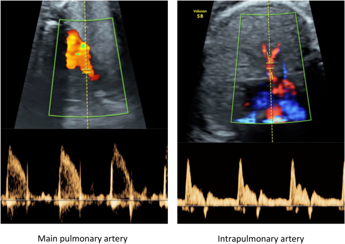
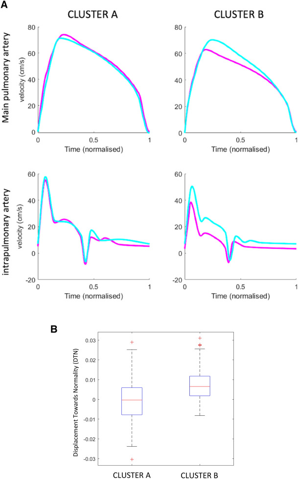
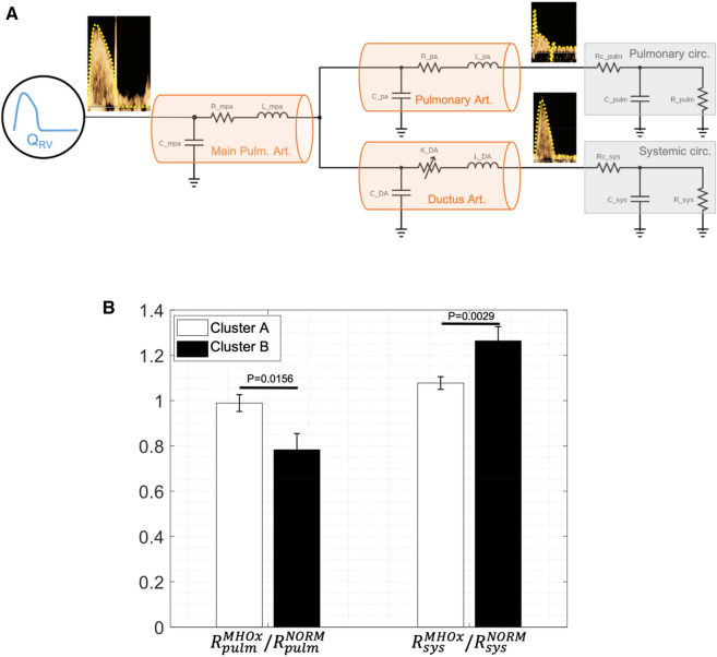
The aim of this study was to investigate the pulmonary vasculature in baseline conditions and after maternal hyperoxygenation in growth restricted fetuses (FGR). A prospective cohort study of singleton pregnancies including 97 FGR and 111 normally grown fetuses was carried out. Ultrasound Doppler of the pulmonary vessels was obtained at 24-37 weeks of gestation and data were acquired before and after oxygen administration. After, Machine Learning (ML) and a computational model were used on the Doppler waveforms to classify individuals and estimate pulmonary vascular resistance (PVR). Our results showed lower mean velocity time integral (VTI) in the main pulmonary and intrapulmonary arteries in baseline conditions in FGR individuals. Delta changes of the main pulmonary artery VTI and intrapulmonary artery pulsatility index before and after hyperoxygenation were significantly greater in FGR when compared with controls. Also, ML identified two clusters: A (including 66% controls and 34% FGR) with similar Doppler traces over time and B (including 33% controls and 67% FGR) with changes after hyperoxygenation. The computational model estimated the ratio of PVR before and after maternal hyperoxygenation which was closer to 1 in cluster A (cluster A 0.98 ± 0.33 vs cluster B 0.78 ± 0.28, p = 0.0156). Doppler ultrasound allows the detection of significant changes in pulmonary vasculature in most FGR at baseline, and distinct responses to hyperoxygenation. Future studies are warranted to assess its potential applicability in the clinical management of FGR.
本研究的目的是调查生长受限胎儿(FGR)在基线状态下以及母体高氧治疗后的肺血管系统。对单胎妊娠进行了一项前瞻性队列研究,包括97例FGR胎儿和111例正常生长的胎儿。在妊娠24 - 37周时获取肺血管的超声多普勒数据,并在吸氧前后采集数据。之后,使用机器学习(ML)和一个计算模型对多普勒波形进行分析,以对个体进行分类并估计肺血管阻力(PVR)。我们的结果显示,在基线状态下,FGR个体的主肺动脉和肺内动脉的平均速度时间积分(VTI)较低。与对照组相比,FGR胎儿在高氧治疗前后主肺动脉VTI和肺内动脉搏动指数的变化明显更大。此外,ML识别出两个集群:A集群(包括66%的对照组和34%的FGR),其多普勒轨迹随时间相似;B集群(包括33%的对照组和67%的FGR),在高氧治疗后有变化。计算模型估计了母体高氧治疗前后PVR的比值,A集群更接近1(A集群0.98±0.33 vs B集群0.78±0.28,p = 0.0156)。多普勒超声能够检测出大多数FGR胎儿在基线时肺血管系统的显著变化,以及对高氧治疗的不同反应。未来有必要进行研究,以评估其在FGR临床管理中的潜在适用性。