• 文献检索
  • 文档翻译
  • 深度研究
  • 学术资讯
  • Suppr Zotero 插件Zotero 插件
  • 邀请有礼
  • 套餐&价格
  • 历史记录
应用&插件
Suppr Zotero 插件Zotero 插件浏览器插件Mac 客户端Windows 客户端微信小程序
定价
高级版会员购买积分包购买API积分包
服务
文献检索文档翻译深度研究API 文档MCP 服务
关于我们
关于 Suppr公司介绍联系我们用户协议隐私条款
关注我们

Suppr 超能文献

核心技术专利:CN118964589B侵权必究
粤ICP备2023148730 号-1Suppr @ 2026

文献检索

告别复杂PubMed语法,用中文像聊天一样搜索,搜遍4000万医学文献。AI智能推荐,让科研检索更轻松。

立即免费搜索

文件翻译

保留排版,准确专业,支持PDF/Word/PPT等文件格式,支持 12+语言互译。

免费翻译文档

深度研究

AI帮你快速写综述,25分钟生成高质量综述,智能提取关键信息,辅助科研写作。

立即免费体验

蛋白激酶D通过调节肌丝蛋白的翻译后修饰在维持心脏稳态中起关键作用。

Protein Kinase D Plays a Crucial Role in Maintaining Cardiac Homeostasis by Regulating Post-Translational Modifications of Myofilament Proteins.

作者信息

Herwig Melissa, Begovic Merima, Budde Heidi, Delalat Simin, Zhazykbayeva Saltanat, Sieme Marcel, Schneider Luca, Jaquet Kornelia, Mügge Andreas, Akin Ibrahim, El-Battrawy Ibrahim, Fielitz Jens, Hamdani Nazha

机构信息

Department of Cellular and Translational Physiology, Institute of Physiology, Ruhr University Bochum, 44801 Bochum, Germany.

Institut für Forschung und Lehre (IFL), Molecular and Experimental Cardiology, Ruhr University Bochum, 44791 Bochum, Germany.

出版信息

Int J Mol Sci. 2024 Feb 28;25(5):2790. doi: 10.3390/ijms25052790.

DOI:10.3390/ijms25052790
PMID:38474037
原文链接:https://pmc.ncbi.nlm.nih.gov/articles/PMC10932171/
Abstract

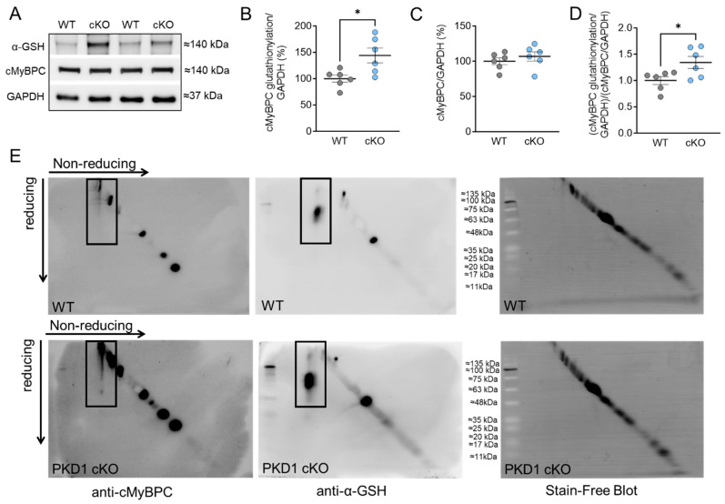
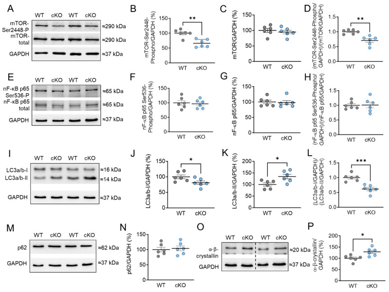
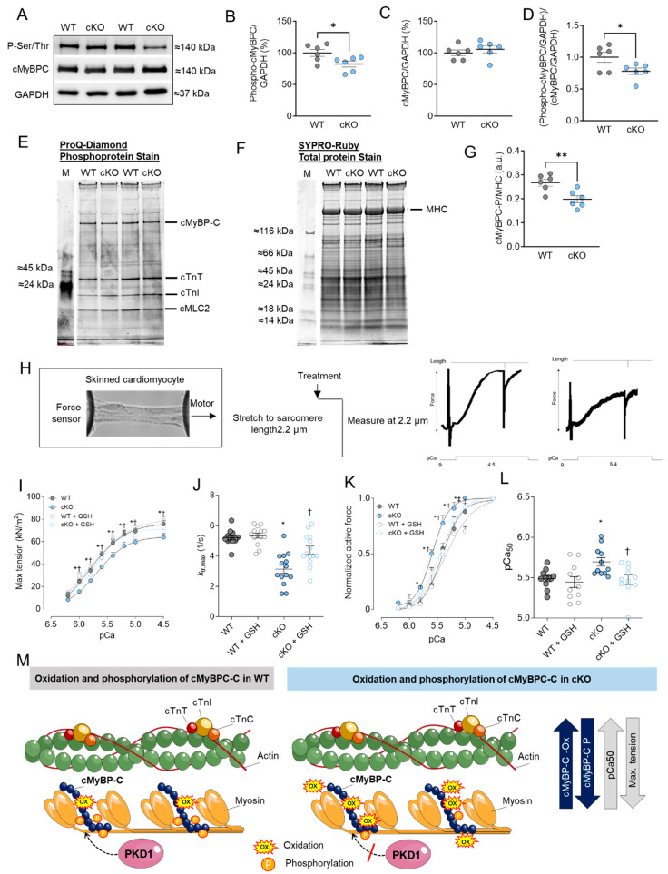
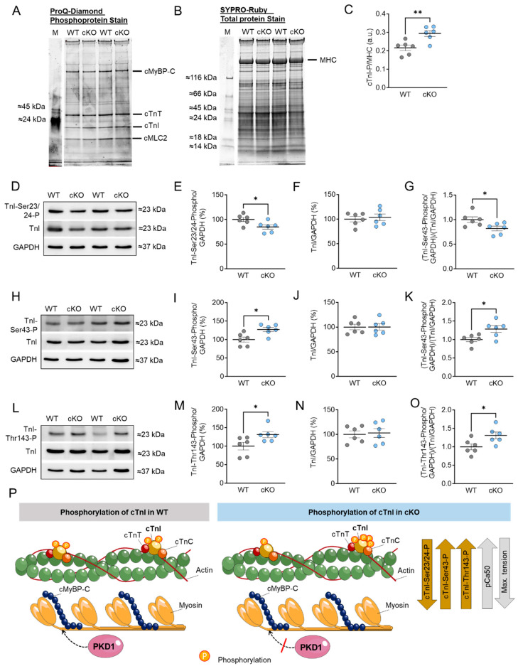
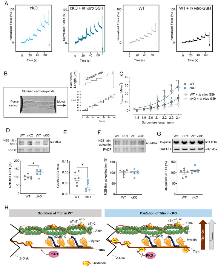
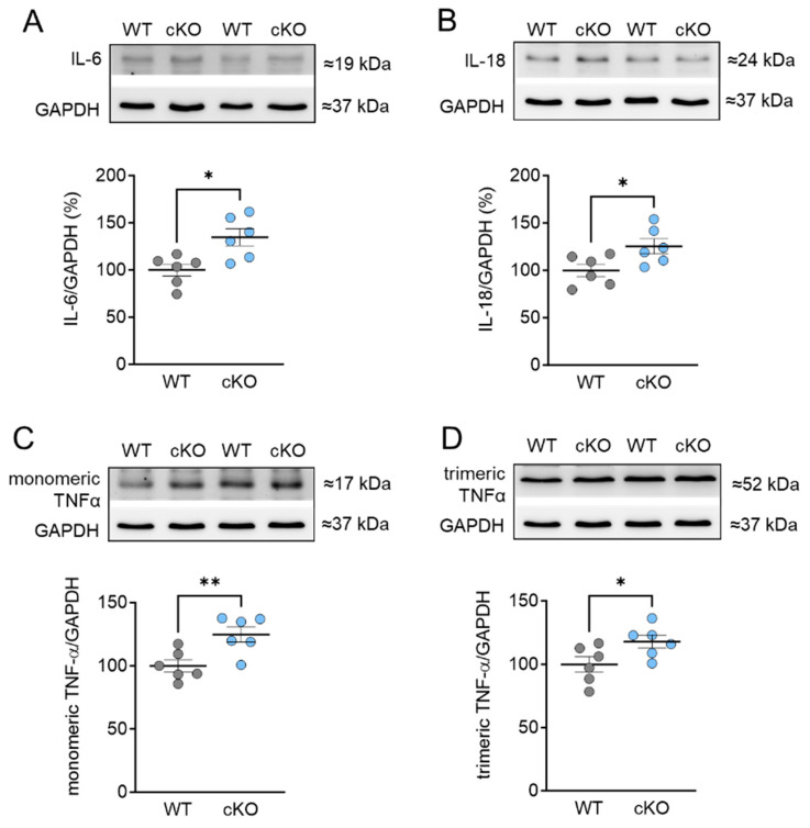
Protein kinase D (PKD) enzymes play important roles in regulating myocardial contraction, hypertrophy, and remodeling. One of the proteins phosphorylated by PKD is titin, which is involved in myofilament function. In this study, we aimed to investigate the role of PKD in cardiomyocyte function under conditions of oxidative stress. To do this, we used mice with a cardiomyocyte-specific knock-out of Prkd1, which encodes PKD1 (Prkd1; ; PKD1 cKO), as well as wild type littermate controls (Prkd1; WT). We isolated permeabilized cardiomyocytes from PKD1 cKO mice and found that they exhibited increased passive stiffness (F), which was associated with increased oxidation of titin, but showed no change in titin ubiquitination. Additionally, the PKD1 cKO mice showed increased myofilament calcium (Ca) sensitivity (pCa) and reduced maximum Ca-activated tension. These changes were accompanied by increased oxidation and reduced phosphorylation of the small myofilament protein cardiac myosin binding protein C (cMyBPC), as well as altered phosphorylation levels at different phosphosites in troponin I (TnI). The increased F and pCa, and the reduced maximum Ca-activated tension were reversed when we treated the isolated permeabilized cardiomyocytes with reduced glutathione (GSH). This indicated that myofilament protein oxidation contributes to cardiomyocyte dysfunction. Furthermore, the PKD1 cKO mice exhibited increased oxidative stress and increased expression of pro-inflammatory markers interleukin (IL)-6, IL-18, and tumor necrosis factor alpha (TNF-α). Both oxidative stress and inflammation contributed to an increase in microtubule-associated protein 1 light chain 3 (LC3)-II levels and heat shock response by inhibiting the mammalian target of rapamycin (mTOR) in the PKD1 cKO mouse myocytes. These findings revealed a previously unknown role for PKD1 in regulating diastolic passive properties, myofilament Ca sensitivity, and maximum Ca-activated tension under conditions of oxidative stress. Finally, we emphasized the importance of PKD1 in maintaining the balance of oxidative stress and inflammation in the context of autophagy, as well as cardiomyocyte function.

摘要

蛋白激酶D(PKD)酶在调节心肌收缩、肥大和重塑中发挥着重要作用。被PKD磷酸化的蛋白之一是肌联蛋白,它参与肌丝功能。在本研究中,我们旨在研究PKD在氧化应激条件下对心肌细胞功能的作用。为此,我们使用了心肌细胞特异性敲除编码PKD1的Prkd1的小鼠(Prkd1;;PKD1 cKO),以及野生型同窝对照(Prkd1;WT)。我们从PKD1 cKO小鼠中分离出透化心肌细胞,发现它们表现出被动僵硬度(F)增加,这与肌联蛋白氧化增加有关,但肌联蛋白泛素化没有变化。此外,PKD1 cKO小鼠表现出肌丝钙(Ca)敏感性(pCa)增加和最大钙激活张力降低。这些变化伴随着小肌丝蛋白心肌肌球蛋白结合蛋白C(cMyBPC)氧化增加和磷酸化减少,以及肌钙蛋白I(TnI)不同磷酸位点的磷酸化水平改变。当我们用还原型谷胱甘肽(GSH)处理分离的透化心肌细胞时,增加的F和pCa以及降低的最大钙激活张力得到逆转。这表明肌丝蛋白氧化导致心肌细胞功能障碍。此外,PKD1 cKO小鼠表现出氧化应激增加以及促炎标志物白细胞介素(IL)-6、IL-18和肿瘤坏死因子α(TNF-α)表达增加。氧化应激和炎症都通过抑制PKD1 cKO小鼠心肌细胞中的雷帕霉素靶蛋白(mTOR)导致微管相关蛋白1轻链3(LC3)-II水平增加和热休克反应。这些发现揭示了PKD1在氧化应激条件下调节舒张期被动特性、肌丝钙敏感性和最大钙激活张力方面以前未知的作用。最后,我们强调了PKD1在自噬背景下维持氧化应激和炎症平衡以及心肌细胞功能方面的重要性。

https://cdn.ncbi.nlm.nih.gov/pmc/blobs/fd0f/10932171/a32afa14de6d/ijms-25-02790-g006.jpg
https://cdn.ncbi.nlm.nih.gov/pmc/blobs/fd0f/10932171/ef852c9292ae/ijms-25-02790-g001.jpg
https://cdn.ncbi.nlm.nih.gov/pmc/blobs/fd0f/10932171/d188513168de/ijms-25-02790-g002.jpg
https://cdn.ncbi.nlm.nih.gov/pmc/blobs/fd0f/10932171/2e3fc24b98ad/ijms-25-02790-g003.jpg
https://cdn.ncbi.nlm.nih.gov/pmc/blobs/fd0f/10932171/0139338b6db7/ijms-25-02790-g004.jpg
https://cdn.ncbi.nlm.nih.gov/pmc/blobs/fd0f/10932171/d633cbff26b6/ijms-25-02790-g005.jpg
https://cdn.ncbi.nlm.nih.gov/pmc/blobs/fd0f/10932171/a32afa14de6d/ijms-25-02790-g006.jpg

相似文献

1
Protein Kinase D Plays a Crucial Role in Maintaining Cardiac Homeostasis by Regulating Post-Translational Modifications of Myofilament Proteins.蛋白激酶D通过调节肌丝蛋白的翻译后修饰在维持心脏稳态中起关键作用。
Int J Mol Sci. 2024 Feb 28;25(5):2790. doi: 10.3390/ijms25052790.
2
Modulation of Titin-Based Stiffness in Hypertrophic Cardiomyopathy via Protein Kinase D.通过蛋白激酶D调节肥厚型心肌病中基于肌联蛋白的僵硬度
Front Physiol. 2020 Apr 15;11:240. doi: 10.3389/fphys.2020.00240. eCollection 2020.
3
Diabetes-Induced Cardiomyocyte Passive Stiffening Is Caused by Impaired Insulin-Dependent Titin Modification and Can Be Modulated by Neuregulin-1.糖尿病诱导的心肌细胞被动僵硬度增加是由于胰岛素依赖性肌联蛋白修饰受损引起的,可通过神经调节素-1进行调节。
Circ Res. 2018 Jul 20;123(3):342-355. doi: 10.1161/CIRCRESAHA.117.312166. Epub 2018 May 14.
4
Deranged myofilament phosphorylation and function in experimental heart failure with preserved ejection fraction.实验性射血分数保留心力衰竭中线粒体肌丝磷酸化和功能障碍。
Cardiovasc Res. 2013 Mar 1;97(3):464-71. doi: 10.1093/cvr/cvs353. Epub 2012 Dec 4.
5
Protein kinase D increases maximal Ca2+-activated tension of cardiomyocyte contraction by phosphorylation of cMyBP-C-Ser315.蛋白激酶 D 通过磷酸化 cMyBP-C-Ser315 增加心肌细胞收缩的最大 Ca2+激活张力。
Am J Physiol Heart Circ Physiol. 2012 Aug 1;303(3):H323-31. doi: 10.1152/ajpheart.00749.2011. Epub 2012 May 25.
6
Protein kinase D is a novel mediator of cardiac troponin I phosphorylation and regulates myofilament function.蛋白激酶D是心肌肌钙蛋白I磷酸化的新型介质,并调节肌丝功能。
Circ Res. 2004 Nov 26;95(11):1091-9. doi: 10.1161/01.RES.0000149299.34793.3c. Epub 2004 Oct 28.
7
GSK-3β Localizes to the Cardiac Z-Disc to Maintain Length Dependent Activation.糖原合成酶激酶3β定位于心脏Z线以维持长度依赖性激活。
Circ Res. 2022 Mar 18;130(6):871-886. doi: 10.1161/CIRCRESAHA.121.319491. Epub 2022 Feb 16.
8
Protein kinase D selectively targets cardiac troponin I and regulates myofilament Ca2+ sensitivity in ventricular myocytes.蛋白激酶D选择性作用于心肌肌钙蛋白I并调节心室肌细胞中肌丝对钙离子的敏感性。
Circ Res. 2007 Mar 30;100(6):864-73. doi: 10.1161/01.RES.0000260809.15393.fa. Epub 2007 Feb 22.
9
Crucial role for Ca2(+)/calmodulin-dependent protein kinase-II in regulating diastolic stress of normal and failing hearts via titin phosphorylation.钙/钙调蛋白依赖性蛋白激酶-II 在调节正常和衰竭心脏舒张压力中的关键作用通过肌联蛋白磷酸化实现。
Circ Res. 2013 Feb 15;112(4):664-74. doi: 10.1161/CIRCRESAHA.111.300105. Epub 2013 Jan 2.
10
Selective phosphorylation of PKA targets after β-adrenergic receptor stimulation impairs myofilament function in Mybpc3-targeted HCM mouse model.β-肾上腺素受体刺激后 PKA 靶标的选择性磷酸化可损害 Mybpc3 靶向 HCM 小鼠模型中的肌丝功能。
Cardiovasc Res. 2016 May 15;110(2):200-14. doi: 10.1093/cvr/cvw026. Epub 2016 Jan 29.

引用本文的文献

1
Dysregulated inflammation, oxidative stress, and protein quality control in diabetic HFpEF: unraveling mechanisms and therapeutic targets.糖尿病性射血分数保留的心力衰竭中炎症失调、氧化应激与蛋白质质量控制:揭示机制与治疗靶点
Cardiovasc Diabetol. 2025 May 14;24(1):211. doi: 10.1186/s12933-025-02734-4.
2
Diabetes mellitus aggravates myocardial inflammation and oxidative stress in aortic stenosis: a mechanistic link to HFpEF features.糖尿病会加重主动脉瓣狭窄中的心肌炎症和氧化应激:与射血分数保留的心力衰竭特征的机制联系。
Cardiovasc Diabetol. 2025 May 13;24(1):203. doi: 10.1186/s12933-025-02748-y.

本文引用的文献

1
Stress activated signalling impaired protein quality control pathways in human hypertrophic cardiomyopathy.应激激活的信号通路损害了人类肥厚型心肌病中的蛋白质质量控制途径。
Int J Cardiol. 2021 Dec 1;344:160-169. doi: 10.1016/j.ijcard.2021.09.009. Epub 2021 Sep 10.
2
The Interplay between S-Glutathionylation and Phosphorylation of Cardiac Troponin I and Myosin Binding Protein C in End-Stage Human Failing Hearts.终末期人类衰竭心脏中心肌肌钙蛋白I和肌球蛋白结合蛋白C的S-谷胱甘肽化与磷酸化之间的相互作用
Antioxidants (Basel). 2021 Jul 16;10(7):1134. doi: 10.3390/antiox10071134.
3
Reactive Oxygen Species: Drivers of Physiological and Pathological Processes.
活性氧:生理和病理过程的驱动因素
J Inflamm Res. 2020 Dec 2;13:1057-1073. doi: 10.2147/JIR.S275595. eCollection 2020.
4
Modulation of Titin-Based Stiffness in Hypertrophic Cardiomyopathy via Protein Kinase D.通过蛋白激酶D调节肥厚型心肌病中基于肌联蛋白的僵硬度
Front Physiol. 2020 Apr 15;11:240. doi: 10.3389/fphys.2020.00240. eCollection 2020.
5
Posttranslational modifications of titin from cardiac muscle: how, where, and what for?肌联蛋白的翻译后修饰:如何、何地及为何发生?
FEBS J. 2019 Jun;286(12):2240-2260. doi: 10.1111/febs.14854. Epub 2019 Apr 29.
6
Autophagy: An Essential Degradation Program for Cellular Homeostasis and Life.自噬:细胞稳态与生命的重要降解程序
Cells. 2018 Dec 19;7(12):278. doi: 10.3390/cells7120278.
7
Diagonal Electrophoresis for the Detection of Proteins Involved in Disulfide Bonds.用于检测参与二硫键的蛋白质的对角线电泳
Methods Mol Biol. 2019;1855:279-286. doi: 10.1007/978-1-4939-8793-1_23.
8
Stress-induced protein S-glutathionylation and phosphorylation crosstalk in cardiac sarcomeric proteins - Impact on heart function.应激诱导的心肌肌球蛋白蛋白 S-谷胱甘肽化和磷酸化相互作用——对心脏功能的影响。
Int J Cardiol. 2018 May 1;258:207-216. doi: 10.1016/j.ijcard.2017.12.004.
9
Disulfide isomerization reactions in titin immunoglobulin domains enable a mode of protein elasticity.titin 免疫球蛋白结构域中的二硫键异构反应使蛋白质具有弹性模式。
Nat Commun. 2018 Jan 12;9(1):185. doi: 10.1038/s41467-017-02528-7.
10
PKD knockdown inhibits pressure overload-induced cardiac hypertrophy by promoting autophagy via AKT/mTOR pathway.多囊肾病基因敲低通过AKT/哺乳动物雷帕霉素靶蛋白(mTOR)信号通路促进自噬,从而抑制压力超负荷诱导的心肌肥大。
Int J Biol Sci. 2017 Feb 12;13(3):276-285. doi: 10.7150/ijbs.17617. eCollection 2017.