• 文献检索
  • 文档翻译
  • 深度研究
  • 学术资讯
  • Suppr Zotero 插件Zotero 插件
  • 邀请有礼
  • 套餐&价格
  • 历史记录
应用&插件
Suppr Zotero 插件Zotero 插件浏览器插件Mac 客户端Windows 客户端微信小程序
定价
高级版会员购买积分包购买API积分包
服务
文献检索文档翻译深度研究API 文档MCP 服务
关于我们
关于 Suppr公司介绍联系我们用户协议隐私条款
关注我们

Suppr 超能文献

核心技术专利:CN118964589B侵权必究
粤ICP备2023148730 号-1Suppr @ 2026

文献检索

告别复杂PubMed语法,用中文像聊天一样搜索,搜遍4000万医学文献。AI智能推荐,让科研检索更轻松。

立即免费搜索

文件翻译

保留排版,准确专业,支持PDF/Word/PPT等文件格式,支持 12+语言互译。

免费翻译文档

深度研究

AI帮你快速写综述,25分钟生成高质量综述,智能提取关键信息,辅助科研写作。

立即免费体验

成像模态在黑色素瘤诊断中的应用:一项范围综述

Utilization of Imaging Modalities in the Diagnosis of Melanoma: A Scoping Review.

作者信息

Shapiro Lucas, Basra Mahi, Patel Hemangi, Payne Collin, Brazen Brett, Biglione Alejandro

机构信息

Osteopathic Medicine, Nova Southeastern University Dr. Kiran C. Patel College of Osteopathic Medicine, Clearwater, USA.

Sports Medicine, Nova Southeastern University Dr. Kiran C. Patel College of Osteopathic Medicine, Fort Lauderdale, USA.

出版信息

Cureus. 2024 Feb 12;16(2):e54058. doi: 10.7759/cureus.54058. eCollection 2024 Feb.

DOI:10.7759/cureus.54058
PMID:38481925
原文链接:https://pmc.ncbi.nlm.nih.gov/articles/PMC10933824/
Abstract

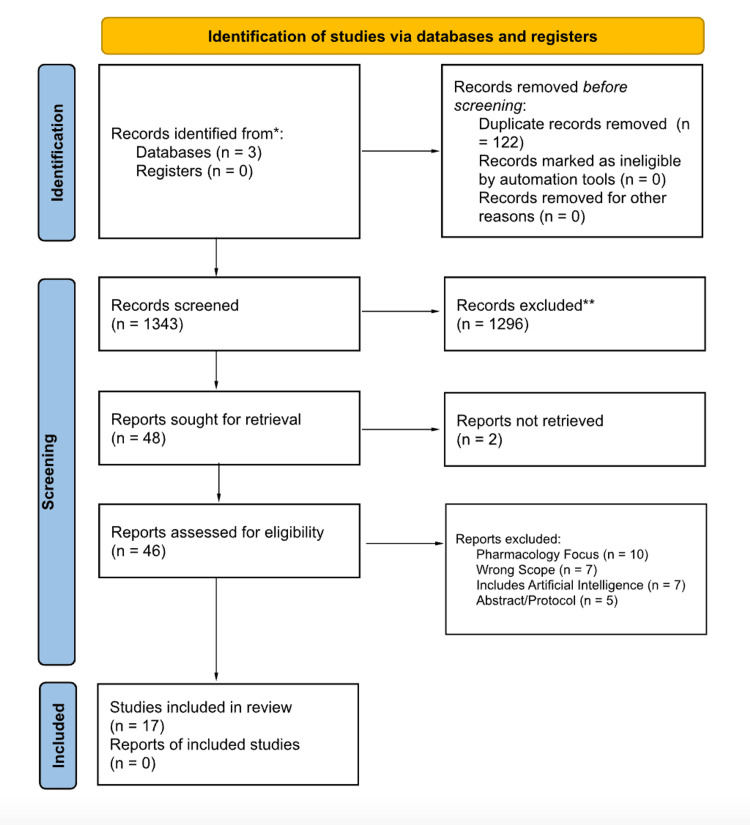
Melanomas arise de novo or in the context of a precursor lesion. Lesions typically grow radially and then undergo a vertical growth phase proceeding to local invasion and metastasis. This review describes the utility of different imaging modalities in diagnosis and melanocytic lesion monitoring. A literature search was performed in November 2023 utilizing EMBASE, Medline, and PubMed. The PRISMA diagram demonstrates the review process. Reflectance confocal microscopy (RCM) utilizes near-infrared light to help diagnose dermatologic lesions. RCM was found to demonstrate nearly two times the positive predictive value compared to dermoscopy. The introduction of the Berlin Ultrasound (US) Morphology Criteria permitted a 65-80% improvement in diagnostic sensitivity. US with fine-needle aspiration cytology (FNAC) accurately predicts the necessity for sentinel lymph node biopsy and lymphadenectomy, sparing patients with metastasis and prompting biopsy for equivocal lesions. Single-photon emission computed tomography/computed tomography (SPECT/CT) is an adjunctive tool to anatomically and functionally assess lymphatic invasion. SPECT/CT improves the detection of sentinel nodes while decreasing operating time and improving cosmetic outcomes. 18F-fluorodeoxyglucose (18F-FDG) positron emission tomography/computed tomography (PET/CT) with small voxel reconstruction demonstrated increased specificity and sensitivity for detecting in-transit metastases of melanomas, specifically in the limbs. Dermoscopy allows providers to cost-effectively recognize common lesion patterns. Multiphoton microscopy assigns a weight-based score based on malignant features. Optical coherence angiography captures images of vessels to help diagnose equivocal lesions. Utilization of imaging techniques may increase diagnostic accuracy, reduce unnecessary procedures, and help guide treatment plans. Additional research is needed to further characterize the utility of these techniques in order to improve the diagnosis and treatment of melanomas.

摘要

黑色素瘤可原发产生,也可在前驱病变的基础上发生。病变通常先呈放射状生长,然后进入垂直生长阶段,进而发生局部侵袭和转移。本综述描述了不同成像方式在黑色素细胞病变诊断和监测中的应用。2023年11月利用EMBASE、Medline和PubMed进行了文献检索。PRISMA流程图展示了综述过程。反射式共聚焦显微镜(RCM)利用近红外光辅助诊断皮肤病变。研究发现,与皮肤镜检查相比,RCM的阳性预测值几乎高出两倍。柏林超声(US)形态学标准的引入使诊断敏感性提高了65 - 80%。超声联合细针穿刺细胞学检查(FNAC)能准确预测前哨淋巴结活检和淋巴结清扫的必要性,使有转移的患者避免不必要的手术,并促使对可疑病变进行活检。单光子发射计算机断层扫描/计算机断层扫描(SPECT/CT)是一种从解剖学和功能上评估淋巴管侵袭的辅助工具。SPECT/CT提高了前哨淋巴结的检出率,同时减少了手术时间并改善了美容效果。18F - 氟脱氧葡萄糖(18F - FDG)正电子发射断层扫描/计算机断层扫描(PET/CT)结合小体素重建对黑色素瘤的远处转移,尤其是四肢的远处转移,显示出更高的特异性和敏感性。皮肤镜检查使医生能够经济高效地识别常见病变模式。多光子显微镜根据恶性特征赋予基于权重的评分。光学相干血管造影术可采集血管图像以辅助诊断可疑病变。成像技术的应用可能会提高诊断准确性,减少不必要的程序,并有助于指导治疗方案。需要进一步研究以更全面地描述这些技术的效用,从而改善黑色素瘤的诊断和治疗。

https://cdn.ncbi.nlm.nih.gov/pmc/blobs/05e6/10933824/8c0a7d42b348/cureus-0016-00000054058-i01.jpg

相似文献

1
Utilization of Imaging Modalities in the Diagnosis of Melanoma: A Scoping Review.成像模态在黑色素瘤诊断中的应用:一项范围综述
Cureus. 2024 Feb 12;16(2):e54058. doi: 10.7759/cureus.54058. eCollection 2024 Feb.
2
Role of In Vivo Reflectance Confocal Microscopy in the Analysis of Melanocytic Lesions.体内反射共聚焦显微镜在黑素细胞性病变分析中的作用
Acta Dermatovenerol Croat. 2018 Apr;26(1):64-67.
3
Ultrasound, CT, MRI, or PET-CT for staging and re-staging of adults with cutaneous melanoma.超声、CT、MRI或PET-CT用于成人皮肤黑色素瘤的分期及再分期。
Cochrane Database Syst Rev. 2019 Jul 1;7(7):CD012806. doi: 10.1002/14651858.CD012806.pub2.
4
Reflectance confocal microscopy for diagnosing cutaneous melanoma in adults.反射式共聚焦显微镜在成人皮肤黑色素瘤诊断中的应用
Cochrane Database Syst Rev. 2018 Dec 4;12(12):CD013190. doi: 10.1002/14651858.CD013190.
5
Can integrated 18F-FDG PET/MR replace sentinel lymph node resection in malignant melanoma?18F-FDG PET/MR 融合显像能否替代恶性黑色素瘤前哨淋巴结切除术?
Eur J Nucl Med Mol Imaging. 2018 Nov;45(12):2093-2102. doi: 10.1007/s00259-018-4061-9. Epub 2018 Jun 6.
6
More advantages in detecting bone and soft tissue metastases from prostate cancer using F-PSMA PET/CT.使用F-PSMA PET/CT检测前列腺癌骨和软组织转移方面有更多优势。
Hell J Nucl Med. 2019 Jan-Apr;22(1):6-9. doi: 10.1967/s002449910952. Epub 2019 Mar 7.
7
123I-MIBG scintigraphy and 18F-FDG-PET imaging for diagnosing neuroblastoma.用于诊断神经母细胞瘤的123I-间碘苄胍闪烁扫描术和18F-氟代脱氧葡萄糖正电子发射断层显像
Cochrane Database Syst Rev. 2015 Sep 29;2015(9):CD009263. doi: 10.1002/14651858.CD009263.pub2.
8
Dermoscopy, with and without visual inspection, for diagnosing melanoma in adults.使用或不使用肉眼检查的皮肤镜检查在成人黑色素瘤诊断中的应用
Cochrane Database Syst Rev. 2018 Dec 4;12(12):CD011902. doi: 10.1002/14651858.CD011902.pub2.
9
Reflectance confocal microscopy for diagnosing keratinocyte skin cancers in adults.成人角质形成细胞皮肤癌诊断中的反射式共聚焦显微镜检查
Cochrane Database Syst Rev. 2018 Dec 4;12(12):CD013191. doi: 10.1002/14651858.CD013191.
10
Reflectance confocal microscopy to diagnose malignant melanoma and lentigo maligna in the UK: a single-centre prospective observational trial.反射式共聚焦显微镜在英国诊断恶性黑色素瘤和恶性雀斑样痣:一项单中心前瞻性观察性试验。
Br J Dermatol. 2024 Dec 23;192(1):27-35. doi: 10.1093/bjd/ljae354.

引用本文的文献

1
Review of Non-Invasive Imaging Technologies for Cutaneous Melanoma.皮肤黑色素瘤的非侵入性成像技术综述
Biosensors (Basel). 2025 May 7;15(5):297. doi: 10.3390/bios15050297.
2
Dysphagia in an 80-Year-Old Woman: A Rare Case of Metastatic Melanoma Presenting in the Small Bowel.一名80岁女性的吞咽困难:小肠转移性黑色素瘤的罕见病例。
Cureus. 2024 Oct 13;16(10):e71373. doi: 10.7759/cureus.71373. eCollection 2024 Oct.
3
Preoperative bimodal imaging evaluation in finding histological correlations of , superficial spreading and nodular melanoma.

本文引用的文献

1
Effect of Reflectance Confocal Microscopy for Suspect Lesions on Diagnostic Accuracy in Melanoma: A Randomized Clinical Trial.反射共聚焦显微镜检查可疑病变对黑素瘤诊断准确性的影响:一项随机临床试验。
JAMA Dermatol. 2022 Jul 1;158(7):754-761. doi: 10.1001/jamadermatol.2022.1570.
2
BRAF V600K vs. BRAF V600E: a comparison of clinical and dermoscopic characteristics and response to immunotherapies and targeted therapies.BRAF V600K 与 BRAF V600E:临床和皮肤镜特征以及对免疫治疗和靶向治疗反应的比较。
Clin Exp Dermatol. 2022 Jun;47(6):1131-1136. doi: 10.1111/ced.15113. Epub 2022 Mar 11.
3
Revisiting detection of in-transit metastases in melanoma patients using digital F-FDG PET/CT with small-voxel reconstruction.
术前双模态成像评估在发现浅表扩散型和结节型黑色素瘤的组织学相关性方面的应用
Front Med (Lausanne). 2024 Aug 9;11:1436078. doi: 10.3389/fmed.2024.1436078. eCollection 2024.
探讨使用小体素重建的数字化 F-FDG PET/CT 检测黑色素瘤患者转移灶。
Ann Nucl Med. 2021 Jun;35(6):669-679. doi: 10.1007/s12149-021-01608-5. Epub 2021 Mar 26.
4
Super-high magnification dermoscopy can aid the differential diagnosis between melanoma and atypical naevi.超高倍皮肤镜检查有助于鉴别黑色素瘤与非典型痣。
Clin Exp Dermatol. 2021 Oct;46(7):1216-1222. doi: 10.1111/ced.14566. Epub 2021 Jul 9.
5
In vivo multimodal optical imaging of dermoscopic equivocal melanocytic skin lesions.皮肤镜下可疑黑素细胞皮肤病变的体内多模态光学成像。
Sci Rep. 2021 Jan 14;11(1):1405. doi: 10.1038/s41598-020-80744-w.
6
Incorporation of dermoscopy improves inter-observer agreement among dermatopathologists in histologic assessment of melanocytic neoplasms.皮肤镜检查的纳入提高了皮肤科病理学家在评估黑色素细胞肿瘤的组织学检查中的观察者间一致性。
Arch Dermatol Res. 2021 Mar;313(2):101-108. doi: 10.1007/s00403-020-02079-w. Epub 2020 Apr 27.
7
Dermoscopic similarity is an independent predictor of BRAF mutational concordance in multiple melanomas.皮肤镜表现相似性是多个黑色素瘤 BRAF 突变一致性的独立预测因子。
Exp Dermatol. 2019 Jul;28(7):829-835. doi: 10.1111/exd.13951. Epub 2019 May 10.
8
Sentinel lymph node excision with or without preoperative hybrid single-photon emission computed tomography/computed tomography (SPECT/CT) in melanoma: study protocol for a multicentric randomized controlled trial.黑色素瘤前哨淋巴结切除加或不加术前混合单光子发射计算机断层扫描/计算机断层扫描(SPECT/CT):一项多中心随机对照试验的研究方案
Trials. 2019 Feb 4;20(1):99. doi: 10.1186/s13063-019-3197-7.
9
Long-term results of ultrasound guided fine needle aspiration cytology in conjunction with sentinel node biopsy support step-wise approach in melanoma.超声引导下细针穿刺细胞学检查联合前哨淋巴结活检在黑色素瘤中的长期结果支持逐步治疗方法。
Eur J Surg Oncol. 2017 Aug;43(8):1509-1516. doi: 10.1016/j.ejso.2017.02.009. Epub 2017 Feb 21.
10
Paired comparison of the sensitivity and specificity of multispectral digital skin lesion analysis and reflectance confocal microscopy in the detection of melanoma in vivo: A cross-sectional study.多光谱数字皮肤病变分析与反射共聚焦显微镜检测体内黑色素瘤的敏感性和特异性的配对比较:一项横断面研究。
J Am Acad Dermatol. 2016 Dec;75(6):1187-1192.e2. doi: 10.1016/j.jaad.2016.07.022. Epub 2016 Sep 29.