Santos Inês, Simão Maria I, Lúcio Maria J, Santos Miguel O, Sacramento Guilherme, Cabral Catarina I, Vaz Joana
Internal Medicine, Hospital Egas Moniz, Lisbon, PRT.
Gastroenterology, Hospital Egas Moniz, Lisbon, PRT.
Cureus. 2024 Feb 19;16(2):e54446. doi: 10.7759/cureus.54446. eCollection 2024 Feb.
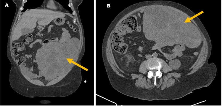
Gastrointestinal stromal tumors (GISTs) arise from the gastrointestinal tract. In rare cases, extra-gastrointestinal stromal tumors (EGISTs) occur in the omentum, mesentery, et cetera. They are mostly asymptomatic or have unspecific symptoms. Risk stratification classification systems are based on tumor size, mitotic rate, location, and perforation. The gold standard for diagnosis is a computed tomography (CT) scan. Ultrasound/CT-guided percutaneous biopsy allows histopathology and immunochemistry results (most stain positive for CD117 (c-KIT), CD34, and/or DOG1). Mutational analysis (most are in proto-oncogene c-KIT and platelet-derived growth factor receptor A (PDGFRA)) determines appropriate therapy. Surgical resection is the gold standard of treatment, with adjuvant and neoadjuvant molecular-targeted therapies depending on recurrence risk and mutations. This report describes a rare case of GIST (omentum EGIST) with a rare presentation (acute pyelonephritis) in a 67-year-old woman. Abdominal examination showed tenderness and a positive Murphy sign on the left side. Blood analysis presented microcytic hypochromic anemia, aggravated renal function, leukocytosis, and increased C-reactive protein. Abdominal CT revealed a heterogeneous abdominal mass, and a CT-guided biopsy showed epithelioid cells positive for CD117 and DOG1, which is compatible with a GIST. The patient underwent surgery that determined the GIST's origin from the greater omentum. Histology revealed an epithelioid GIST with large dimensions and a high histologic grade. Genetic testing detected a variant in the gene. With a high risk of progression, the patient received a three-year course of imatinib.
胃肠道间质瘤(GISTs)起源于胃肠道。在罕见情况下,胃肠道外间质瘤(EGISTs)发生于网膜、肠系膜等部位。它们大多无症状或有非特异性症状。风险分层分类系统基于肿瘤大小、有丝分裂率、位置和穿孔情况。诊断的金标准是计算机断层扫描(CT)。超声/CT引导下经皮活检可得出组织病理学和免疫化学结果(大多数对CD117(c-KIT)、CD34和/或DOG1染色呈阳性)。突变分析(大多数发生在原癌基因c-KIT和血小板衍生生长因子受体A(PDGFRA)中)决定合适的治疗方法。手术切除是治疗的金标准,根据复发风险和突变情况进行辅助和新辅助分子靶向治疗。本报告描述了一名67岁女性罕见的GIST(网膜EGIST)病例,其表现罕见(急性肾盂肾炎)。腹部检查显示左侧有压痛和墨菲氏征阳性。血液分析显示小细胞低色素性贫血、肾功能恶化、白细胞增多和C反应蛋白升高。腹部CT显示腹部有一个不均匀肿块,CT引导下活检显示上皮样细胞CD117和DOG1呈阳性,符合GIST。患者接受了手术,确定GIST起源于大网膜。组织学显示为一个大尺寸、高组织学分级的上皮样GIST。基因检测在该基因中检测到一个变异。由于进展风险高,患者接受了三年的伊马替尼治疗疗程。