Department of Diagnostic and Therapeutic Radiology, Mahidol University, Bangkok, 10400, Thailand.
Department of Medicine, Mahidol University, Bangkok, 10400, Thailand.
Sci Rep. 2024 Mar 22;14(1):6895. doi: 10.1038/s41598-024-57324-3.
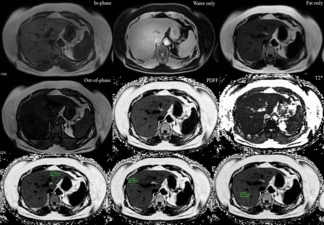
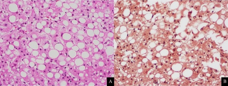
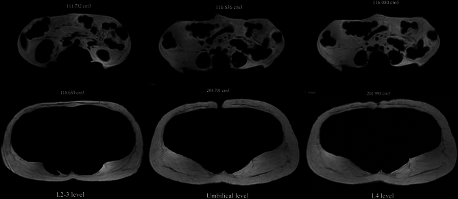
Obesity is highly associated with Non-alcoholic fatty liver disease (NAFLD) and increased risk of liver cirrhosis and liver cancer-related death. We determined the diagnostic performance of the complex-based chemical shift technique MRI-PDFF for quantifying liver fat and its correlation with histopathologic findings in an obese population within 24 h before bariatric surgery. This was a prospective, cross-sectional, Institutional Review Board-approved study of PDFF-MRI of the liver and MRI-DIXON image volume before bariatric surgery. Liver tissues were obtained during bariatric surgery. The prevalence of NAFLD in the investigated cohort was as high as 94%. Histologic hepatic steatosis grades 0, 1, 2, and 3 were observed in 3 (6%), 25 (50%), 14 (28%), and 8 (16%) of 50 obese patients, respectively. The mean percentages of MRI-PDFF from the anterior and posterior right hepatic lobe and left lobe vs. isolate left hepatic lobe were 15.6% (standard deviation [SD], 9.28%) vs. 16.29% (SD, 9.25%). There was a strong correlation between the percentage of steatotic hepatocytes and MRI-PDFF in the left hepatic lobe (r = 0.82, p < 0.001) and the mean value (r = 0.78, p < 0.001). There was a strong correlation between MRI-derived subcutaneous adipose tissue volume and total body fat mass by dual-energy X-ray absorptiometry, especially at the L2-3 and L4 level (r = 0.85, p < 0.001). MRI-PDFF showed good performance in assessing hepatic steatosis and was an excellent noninvasive technique for monitoring hepatic steatosis in an obese population.
肥胖与非酒精性脂肪性肝病(NAFLD)密切相关,并增加肝硬化和肝癌相关死亡的风险。我们在肥胖人群接受减重手术前 24 小时内,通过基于化学位移的磁共振质子密度脂肪分数(MRI-PDFF)技术,评估了该技术对肝脏脂肪的诊断性能及其与组织病理学检查结果的相关性。这是一项前瞻性、横断面、机构审查委员会批准的研究,旨在评估减重手术前肝脏 PDFF-MRI 和 MRI-DIXON 图像体积。在减重手术过程中获取肝脏组织。在所研究的队列中,NAFLD 的患病率高达 94%。50 名肥胖患者中,分别有 3 名(6%)、25 名(50%)、14 名(28%)和 8 名(16%)患者的肝组织学检查结果为肝脂肪变性 0 级、1 级、2 级和 3 级。前右叶、后右叶和左叶的 MRI-PDFF 平均值(标准差[SD])分别为 15.6%(SD,9.28%)和 16.29%(SD,9.25%)。左叶肝脂肪变性细胞百分比与 MRI-PDFF 之间存在很强的相关性(r=0.82,p<0.001),与平均值之间也存在很强的相关性(r=0.78,p<0.001)。MRI 测量的皮下脂肪组织体积与双能 X 射线吸收法测量的全身脂肪质量之间存在很强的相关性,尤其是在 L2-3 和 L4 水平(r=0.85,p<0.001)。MRI-PDFF 在评估肝脂肪变性方面具有良好的性能,是监测肥胖人群肝脂肪变性的一种极好的非侵入性技术。