Key Laboratory of Bioorganic Phosphorus Chemistry and Chemical Biology (Ministry of Education), Department of Chemistry, Tsinghua University, Beijing, 100084, China.
Interdisciplinary Research Center on Biology and Chemistry, Shanghai Institute of Organic Chemistry, Chinese Academy of Sciences, Shanghai, 201210, China.
Nat Commun. 2024 Mar 27;15(1):2677. doi: 10.1038/s41467-024-46898-1.
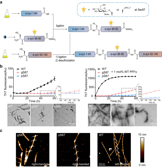
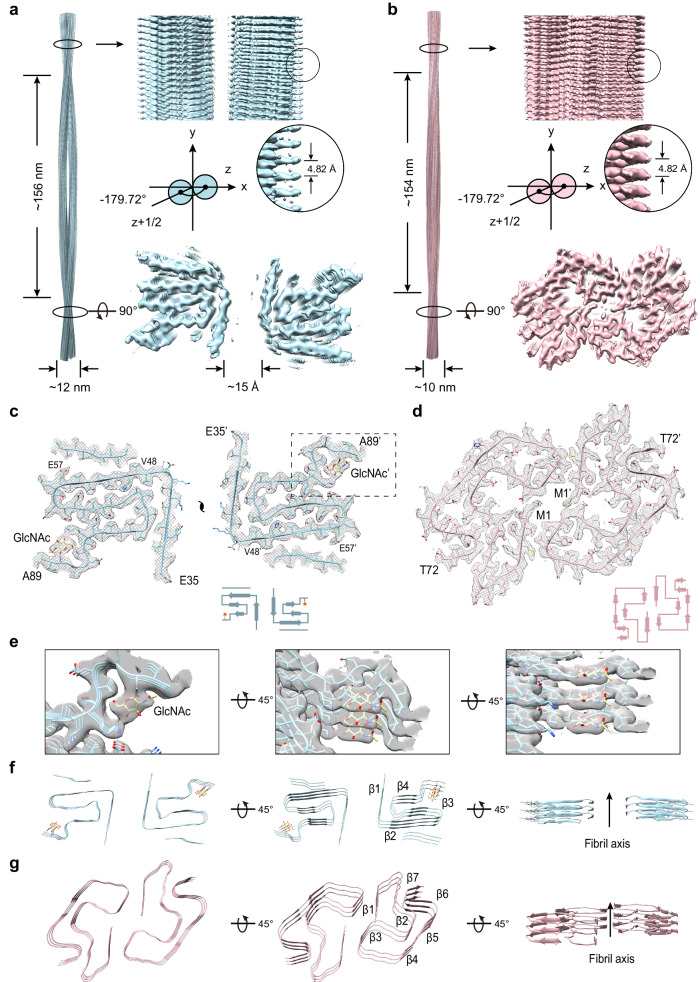
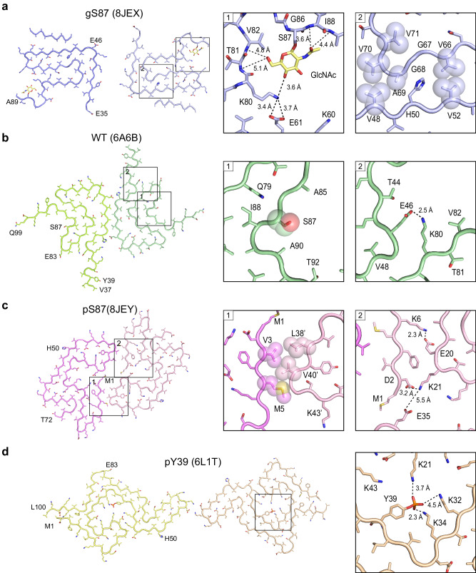
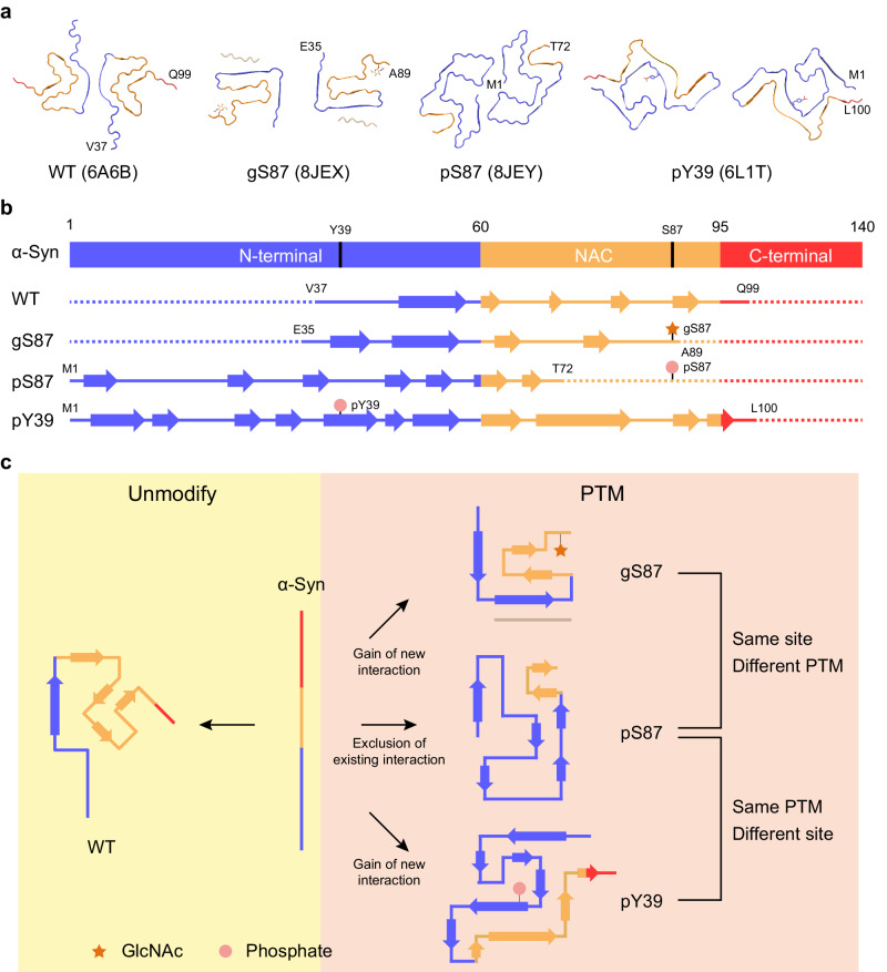
α-Synuclein forms amyloid fibrils that are critical in the progression of Parkinson's disease and serves as the pathological hallmark of this condition. Different posttranslational modifications have been identified at multiple sites of α-synuclein, influencing its conformation, aggregation and function. Here, we investigate how disease-related phosphorylation and O-GlcNAcylation at the same α-synuclein site (S87) affect fibril structure and neuropathology. Using semi-synthesis, we obtained homogenous α-synuclein monomer with site-specific phosphorylation (pS87) and O-GlcNAcylation (gS87) at S87, respectively. Cryo-EM revealed that pS87 and gS87 α-synuclein form two distinct fibril structures. The GlcNAc situated at S87 establishes interactions with K80 and E61, inducing a unique iron-like fold with the GlcNAc molecule on the iron handle. Phosphorylation at the same site prevents a lengthy C-terminal region including residues 73 to 140 from incorporating into the fibril core due to electrostatic repulsion. Instead, the N-terminal half of the fibril (1-72) takes on an arch-like fibril structure. We further show that both pS87 and gS87 α-synuclein fibrils display reduced neurotoxicity and propagation activity compared with unmodified α-synuclein fibrils. Our findings demonstrate that different posttranslational modifications at the same site can produce distinct fibril structures, which emphasizes link between posttranslational modifications and amyloid fibril formation and pathology.
α-突触核蛋白形成的淀粉样纤维在帕金森病的进展中起着关键作用,是这种疾病的病理标志。在α-突触核蛋白的多个位点已经鉴定出不同的翻译后修饰,影响其构象、聚集和功能。在这里,我们研究了疾病相关的磷酸化和 O-GlcNAc 化在同一α-突触核蛋白位点(S87)如何影响纤维结构和神经病理学。通过半合成,我们获得了具有位点特异性磷酸化(pS87)和 O-GlcNAc 化(gS87)的均一的α-突触核蛋白单体。低温电子显微镜揭示了 pS87 和 gS87α-突触核蛋白形成两种不同的纤维结构。位于 S87 处的 GlcNAc 与 K80 和 E61 建立相互作用,诱导 GlcNAc 分子位于铁手柄上的独特铁样折叠。由于静电排斥,同一位置的磷酸化阻止了包括残基 73 到 140 在内的长 C 末端区域掺入纤维核心。相反,纤维的 N 端半部分(1-72)呈现出拱形纤维结构。我们进一步表明,与未修饰的α-突触核蛋白纤维相比,pS87 和 gS87α-突触核蛋白纤维的神经毒性和传播活性均降低。我们的研究结果表明,同一位置的不同翻译后修饰可以产生不同的纤维结构,这强调了翻译后修饰与淀粉样纤维形成和病理学之间的联系。