Hatano Keisuke, Sato Keishiro, Nakamura Tomohiro, Hotta Ryuya, Numoto Shingo, Fujimoto Ayataka
Comprehensive Epilepsy Center, Seirei Hamamatsu General Hospital, 2-12-12 Sumiyoshi, Nakaku, Hamamatsu, Shizuoka, 430-8558, Japan.
Department of Neurosurgery, Seirei Hamamatsu General Hospital, 2-12-12 Sumiyoshi, Nakaku, Hamamatsu, Shizuoka, 430-8558, Japan.
Heliyon. 2024 Mar 18;10(6):e28273. doi: 10.1016/j.heliyon.2024.e28273. eCollection 2024 Mar 30.
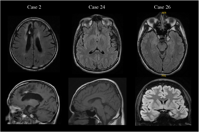
Cavernous malformation (CM) is a well-known cause of epilepsy. Although the location of the CM is usually consistent with the side of seizure onset, some reports have described discrepancies between results from scalp electroencephalography (EEG) and CM location. This study investigated the prevalence and features of patients showing false lateralization (FL). Particularly, we tested the hypothesis that patients showing FL were more likely to have CM in medial and deep areas of the brain than in other areas.
Patients diagnosed with CM-associated epilepsy in our institution between March 2009 and March 2023 were included in this retrospective analysis. We investigated the presence or absence of FL of interictal epileptiform discharges (IEDs) or ictal discharges against MRI findings or against the true focus as determined from surgical outcomes. We compared the FL group with the non-false-lateralization group (NFL group) to clarify features of CM-associated epilepsy patients showing FL.
Thirty-two epilepsy patients with CM were analyzed. The frequency of FL to MRI was 10.3% for IEDs and 7.7% for ictal discharges, while the frequency of FL to true focus after removal surgery was 10.5% for IEDs and 7.7% for ictal discharges. Regarding the FL of IEDs against MRI findings, the percentage of medial and deep lesions was significantly higher in the FL group (3/3, 100%) than in the NFL group (6/26, 23.1%; p = 0.023). No significant differences in age, sex, seizure type, or size of the CM were seen between groups.
CM-associated epilepsy can also present with FL, particularly if the location of the CM is medial and deep. Caution may be needed in determining the area for resection based solely on scalp EEG findings.
海绵状血管畸形(CM)是癫痫的一个众所周知的病因。尽管CM的位置通常与癫痫发作起始侧一致,但一些报告描述了头皮脑电图(EEG)结果与CM位置之间的差异。本研究调查了出现假定位(FL)的患者的患病率和特征。特别是,我们检验了这样一个假设,即出现FL的患者大脑内侧和深部区域的CM比其他区域更常见。
本回顾性分析纳入了2009年3月至2023年3月期间在我们机构被诊断为CM相关性癫痫的患者。我们根据MRI结果或手术结果确定的真正病灶,调查了发作间期癫痫样放电(IEDs)或发作期放电是否存在FL。我们将FL组与非假定位组(NFL组)进行比较,以阐明出现FL的CM相关性癫痫患者的特征。
分析了32例患有CM的癫痫患者。IEDs对MRI的FL发生率为10.3%,发作期放电为7.7%,而切除手术后IEDs对真正病灶的FL发生率为10.5%,发作期放电为7.7%。关于IEDs相对于MRI结果的FL,FL组内侧和深部病变的百分比(3/3,100%)显著高于NFL组(6/26,23.1%;p = 0.023)。两组之间在年龄、性别、癫痫发作类型或CM大小方面没有显著差异。
CM相关性癫痫也可能出现FL,特别是当CM位于内侧和深部时。仅根据头皮EEG结果确定切除区域时可能需要谨慎。