Ohtsuki Kazuya, Sawada Masahiro, Yoshizaki Wataru, Ishimori Takayoshi, Sawamoto Nobukatsu, Fushimi Yasutaka, Toda Hiroki
1Departments of Neurosurgery.
2Diagnostic Radiology, Medical Research Institute Kitano Hospital, Osaka, Japan; and.
J Neurosurg Case Lessons. 2024 Apr 1;7(14). doi: 10.3171/CASE23709.
The ventral intermediate nucleus (Vim) of the thalamus is a surgical target for treating various types of tremor. Because it is difficult to visualize the Vim using standard magnetic resonance imaging, the structure is usually targeted based on the anterior and posterior commissures. This standard targeting method is practical in most patients but not in those with thalamic asymmetry. The authors examined the usefulness of quantitative susceptibility mapping (QSM) and transformed Vim atlas images to estimate the Vim localization in a patient with tremor and significant thalamic hypertrophy.
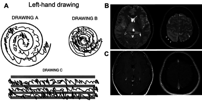
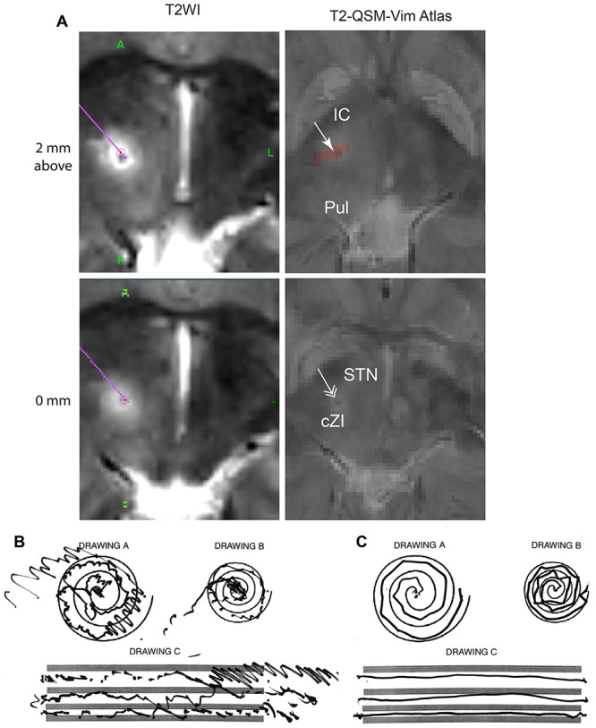
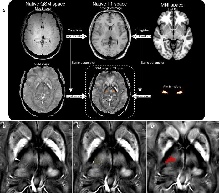
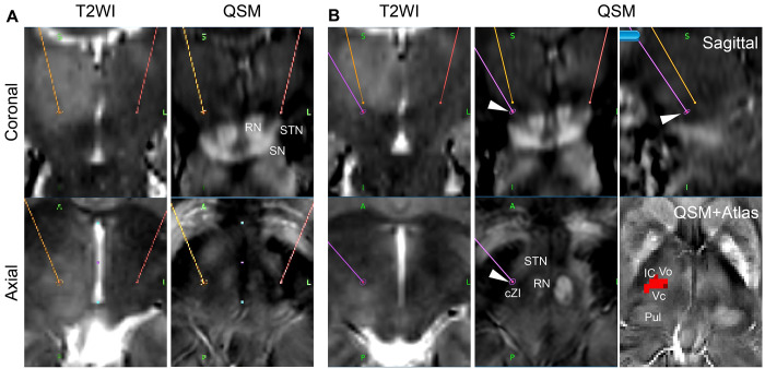
A 51-year-old right-handed female had experienced a predominant left-hand action tremor for 6 years. Magnetic resonance imaging showed significant hypertrophy of the right thalamus and caudal shift of the thalamic ventral border. The authors referred to the QSM images to localize the decreased susceptibility area within the lateral ventral thalamic nuclei to target the Vim. In addition, the nonlinearly transformed Vim atlas images complemented the imaging-based targeting. The radiofrequency thalamotomy at the modified Vim target relieved the tremor completely.
A combination of QSM and nonlinear transformation of the thalamic atlas can be helpful in the targeting method of the Vim for tremor patients with thalamic asymmetry.
丘脑腹中间核(Vim)是治疗各类震颤的手术靶点。由于使用标准磁共振成像难以可视化Vim,该结构通常以前后连合为靶点。这种标准靶向方法在大多数患者中可行,但在丘脑不对称的患者中则不然。作者研究了定量磁化率映射(QSM)和转换后的Vim图谱图像在估计一名患有震颤且丘脑明显肥大患者的Vim定位中的作用。
一名51岁右利手女性有6年以左手为主的动作性震颤病史。磁共振成像显示右侧丘脑明显肥大,丘脑腹侧边界尾侧移位。作者参考QSM图像来定位丘脑外侧腹侧核内磁化率降低的区域,以确定Vim的靶点。此外,非线性转换后的Vim图谱图像补充了基于成像的靶向定位。在改良的Vim靶点进行射频丘脑切开术使震颤完全缓解。
QSM与丘脑图谱的非线性转换相结合,有助于为丘脑不对称的震颤患者确定Vim的靶向方法。