From the Department of Radiology, Balgrist University Hospital, Faculty of Medicine, University of Zurich, Zurich, Switzerland (G.C.F., A.A.M., S.S.G., R.S.); Swiss Center for Musculoskeletal Imaging, Balgrist Campus, Zurich, Switzerland (A.A.M., S.S.); University of Zurich, Institute of Veterinary Pathology, Laboratory for Animal Pathology, Zurich, Switzerland (M.H.); and Advanced Clinical Imaging Technology, Siemens Healthineers International AG, Zurich, Switzerland (S.S.).
Invest Radiol. 2024 Oct 1;59(10):691-698. doi: 10.1097/RLI.0000000000001074. Epub 2024 Apr 10.
The aim of this study was to qualitatively and quantitatively assess changes in bovine flexor tendons before and after collagen degradation and at different angles in relation to the static B 0 field using 3-dimensional ultra-short echo time (UTE) magnetization transfer (MT) imaging within a clinically feasible acquisition time.
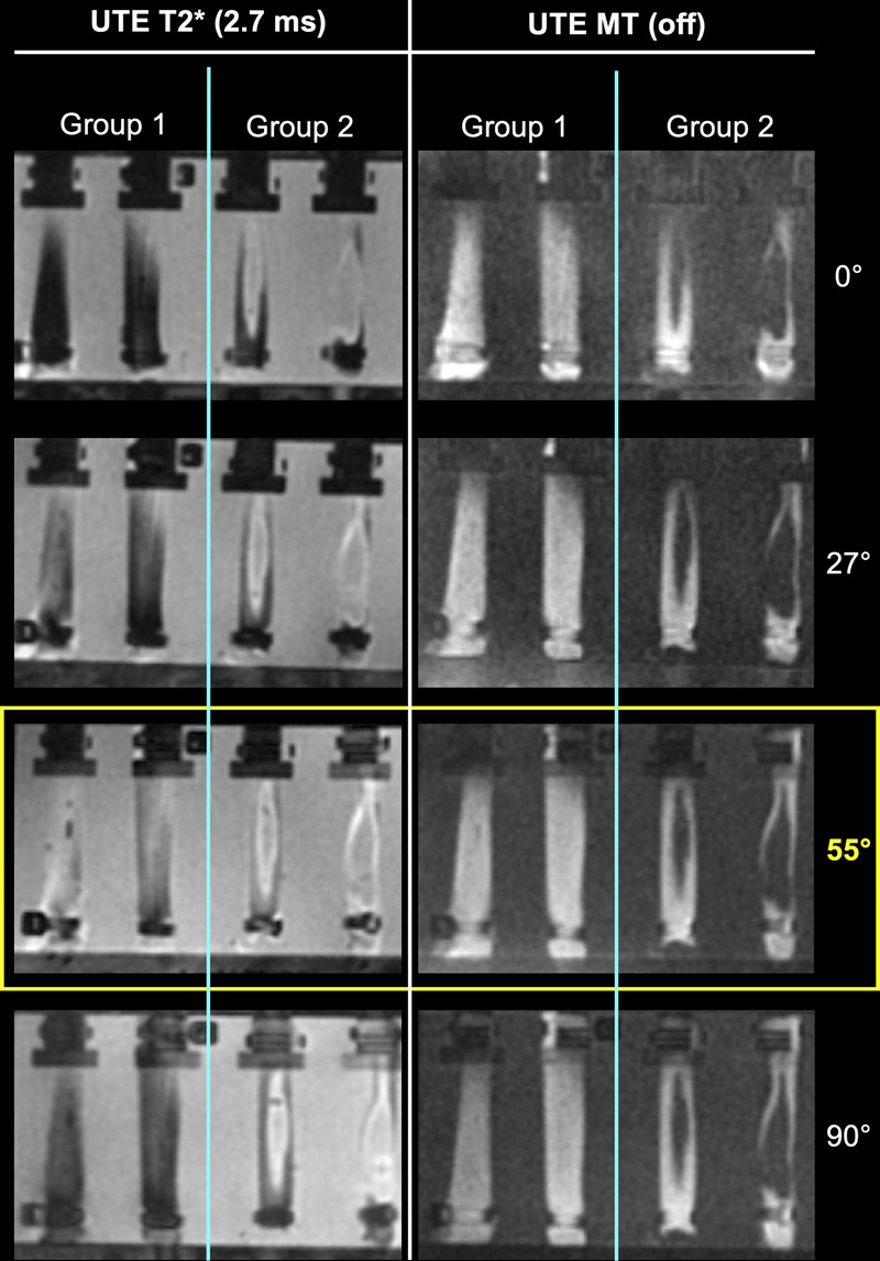
Eight bovine flexor tendons were examined at 3 T magnetic resonance imaging including 3-dimensional UTE MT and UTE T2* research application sequences (acquired within 4:04 and 6:38 minutes, respectively) before and after enzyme-induced degradation. The tendons were divided into 2 groups: group 1 (controls) treated with phosphate-buffered saline and group 2 treated with collagenase I to induce collagen degeneration. Magnetic resonance imaging was repeated at 0, 27, 55, and 90 degrees to the B 0 field. To calculate quantitative tissue properties, all tendons were semiautomatically segmented, and changes in quantitative UTE T2* and UTE MT ratios (MTRs) were compared at different angles and between groups. In addition to descriptive statistics, the coefficient of variation was calculated to compare UTE MT and UTE T2* imaging.
Ultra-short echo time MTR showed a significantly lower coefficient of variation compared with UTE T2* values, indicating a more robust imaging method (UTE MTR 9.64%-11.25%, UTE T2* 18.81%-24.06%, P < 0.001). Both methods showed good performance in detecting degenerated tendons using histopathology as reference standard, with UTE MT imaging having a better area under the curve than UTE T2* mapping (0.918 vs 0.865). Falsely high UTE T2* values were detected at the 55 degrees acquisition angle, whereas UTE MTR values were robust, that is, insensitive to the MAE.
Ultra-short echo time MT imaging is a reliable method for quantifying tendon degeneration that is robust to the MAE and can be acquired in a clinically reasonable time.
本研究旨在使用三维超短回波时间(UTE)磁化传递(MT)成像,在临床可行的采集时间内,定性和定量评估牛屈肌腱在胶原降解前后以及与静态 B0 场不同角度的变化。
在 3T 磁共振成像中检查了 8 个牛屈肌腱,包括三维 UTE MT 和 UTE T2研究应用序列(分别在 4:04 和 6:38 分钟内采集),在酶诱导降解前后。将肌腱分为 2 组:1 组(对照组)用磷酸盐缓冲盐水处理,2 组用胶原酶 I 处理以诱导胶原变性。将磁共振成像重复 0、27、55 和 90 度至 B0 场。为了计算定量组织特性,所有肌腱均进行半自动分割,并比较了不同角度和组间的定量 UTE T2和 UTE MT 比(MTR)的变化。除了描述性统计外,还计算了变异系数以比较 UTE MT 和 UTE T2*成像。
超短回波时间 MTR 的变异系数明显低于 UTE T2值,表明该方法更稳健(UTE MTR 9.64%-11.25%,UTE T2 18.81%-24.06%,P <0.001)。两种方法均使用组织病理学作为参考标准,对变性肌腱的检测性能良好,UTE MT 成像的曲线下面积优于 UTE T2映射(0.918 对 0.865)。在 55 度采集角度检测到错误的高 UTE T2值,而 UTE MTR 值则稳健,即对 MAE 不敏感。
超短回波时间 MT 成像可用于定量评估肌腱变性,该方法稳健,不受 MAE 影响,可在临床合理时间内采集。