Angst Ueli M, Rossi Emanuele, Boschmann Käthler Carolina, Mannes David, Trtik Pavel, Elsener Bernhard, Zhou Zhou, Strobl Markus
Institute for Building Materials, ETH Zurich, Zurich, Switzerland.
Hagerbach Test Gallery Ltd., VSH, Flums, Switzerland.
Mater Struct. 2024;57(4):56. doi: 10.1617/s11527-024-02337-7. Epub 2024 Apr 8.
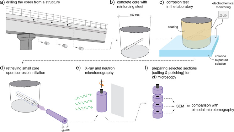
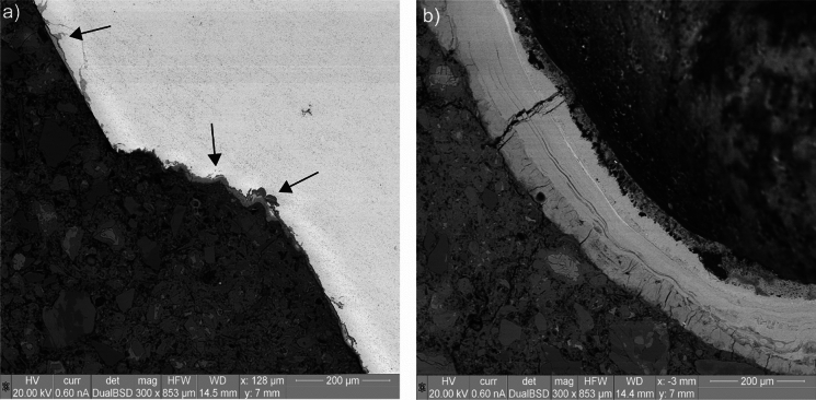
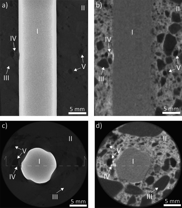
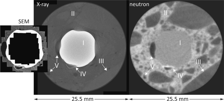
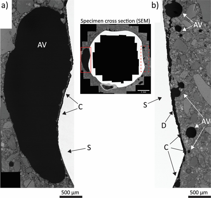
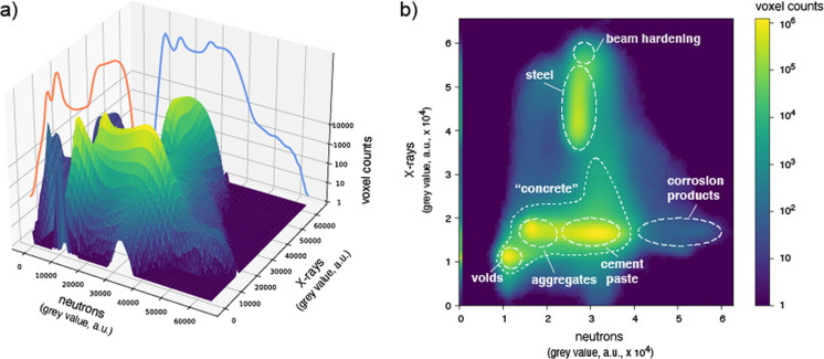
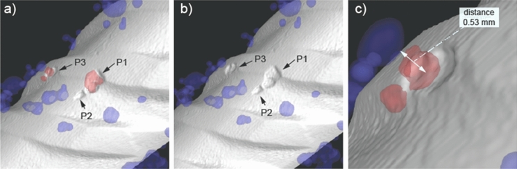
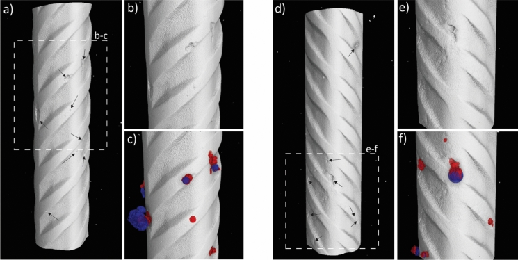
The steel-concrete interface (SCI) is known to play a major role in corrosion of steel in concrete, but a fundamental understanding is still lacking. One reason is that concrete's opacity complicates the study of internal processes. Here, we report on the application of bimodal X-ray and neutron microtomography as in-situ imaging techniques to elucidate the mechanism of steel corrosion in concrete. The study demonstrates that the segmentation of the specimen components of relevance-steel, cementitious matrix, aggregates, voids, corrosion products-obtained through bimodal X-ray and neutron imaging is more reliable than that based on the results of each of the two techniques separately. Further, we suggest the combination of tomographic in-situ imaging with ex-situ SEM analysis of targeted sections, selected based on the segmented tomograms. These in-situ and ex-situ characterization techniques were applied to study localized corrosion in a very early stage under laboratory chloride-exposure conditions, using reinforced concrete cores retrieved from a concrete bridge. Several interesting observations were made. First, the acquired images revealed the formation of several corrosion sites close to each other. Second, the morphology of the corrosion pits was relatively shallow. Finally, only about half of the total 31 corrosion initiation spots were in close proximity to interfacial macroscopic air voids, and > 90% of the more than 160 interfacial macroscopic air voids were free from corrosion. The findings have implications for the mechanistic understanding of corrosion of steel in concrete and suggest that multimodal in-situ imaging is a valuable technique for further related studies.
The online version contains supplementary material available at 10.1617/s11527-024-02337-7.
众所周知,钢 - 混凝土界面(SCI)在混凝土中钢筋的腐蚀过程中起主要作用,但仍缺乏基本的认识。原因之一是混凝土的不透明性使内部过程的研究变得复杂。在此,我们报告了双峰X射线和中子显微断层扫描作为原位成像技术在阐明混凝土中钢筋腐蚀机制方面的应用。研究表明,通过双峰X射线和中子成像获得的相关试样成分(钢、胶凝基体、骨料、孔隙、腐蚀产物)的分割比单独基于两种技术各自的结果更可靠。此外,我们建议将断层原位成像与基于分割断层图像选择的目标截面的非原位扫描电子显微镜(SEM)分析相结合。这些原位和非原位表征技术应用于在实验室氯化物暴露条件下研究非常早期的局部腐蚀,使用从一座混凝土桥梁取回的钢筋混凝土芯样。有几个有趣的发现。首先,获取的图像显示了几个彼此靠近的腐蚀位点的形成。其次,腐蚀坑的形态相对较浅。最后,在总共31个腐蚀起始点中,只有大约一半紧邻界面宏观气孔,并且在160多个界面宏观气孔中,超过90%没有腐蚀。这些发现对理解混凝土中钢筋腐蚀的机理具有启示意义,并表明多模态原位成像是进一步相关研究的一种有价值的技术。
在线版本包含可在10.1617/s11527 - 024 - 02337 - 7获取的补充材料。