• 文献检索
  • 文档翻译
  • 深度研究
  • 学术资讯
  • Suppr Zotero 插件Zotero 插件
  • 邀请有礼
  • 套餐&价格
  • 历史记录
应用&插件
Suppr Zotero 插件Zotero 插件浏览器插件Mac 客户端Windows 客户端微信小程序
定价
高级版会员购买积分包购买API积分包
服务
文献检索文档翻译深度研究API 文档MCP 服务
关于我们
关于 Suppr公司介绍联系我们用户协议隐私条款
关注我们

Suppr 超能文献

核心技术专利:CN118964589B侵权必究
粤ICP备2023148730 号-1Suppr @ 2026

文献检索

告别复杂PubMed语法,用中文像聊天一样搜索,搜遍4000万医学文献。AI智能推荐,让科研检索更轻松。

立即免费搜索

文件翻译

保留排版,准确专业,支持PDF/Word/PPT等文件格式,支持 12+语言互译。

免费翻译文档

深度研究

AI帮你快速写综述,25分钟生成高质量综述,智能提取关键信息,辅助科研写作。

立即免费体验

人类中心体组装的分子结构的时间序列重建。

Time-series reconstruction of the molecular architecture of human centriole assembly.

机构信息

University of Geneva, Department of Molecular and Cellular Biology, Faculty of Sciences, Geneva, Switzerland.

University of Geneva, Department of Genetic and evolution, Faculty of Sciences, Geneva, Switzerland.

出版信息

Cell. 2024 Apr 25;187(9):2158-2174.e19. doi: 10.1016/j.cell.2024.03.025. Epub 2024 Apr 10.

DOI:10.1016/j.cell.2024.03.025
PMID:38604175
原文链接:https://pmc.ncbi.nlm.nih.gov/articles/PMC11060037/
Abstract

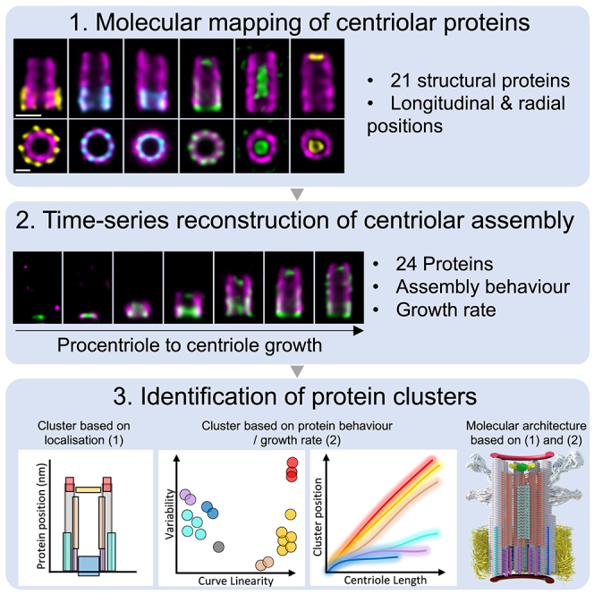
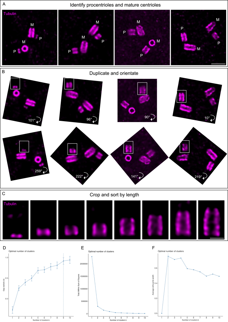
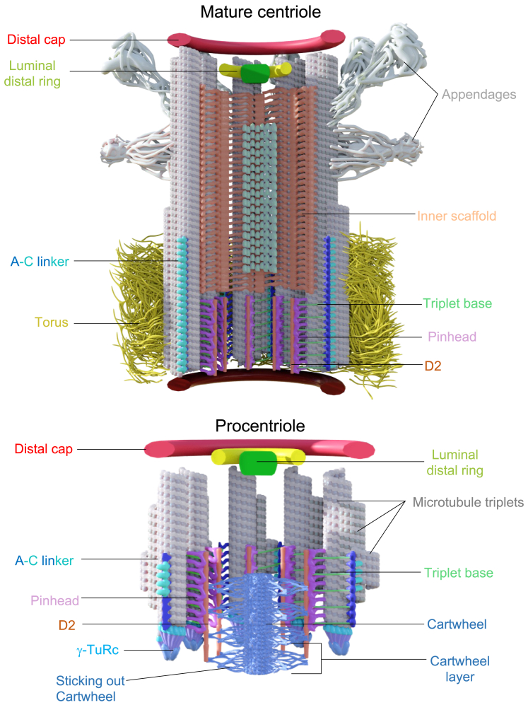
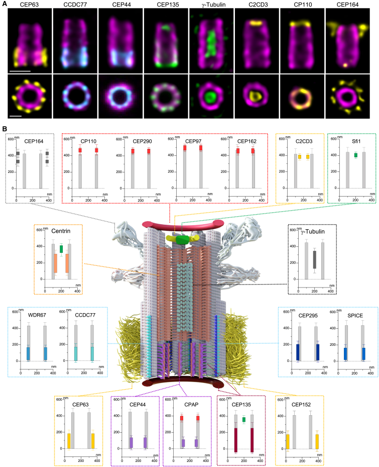
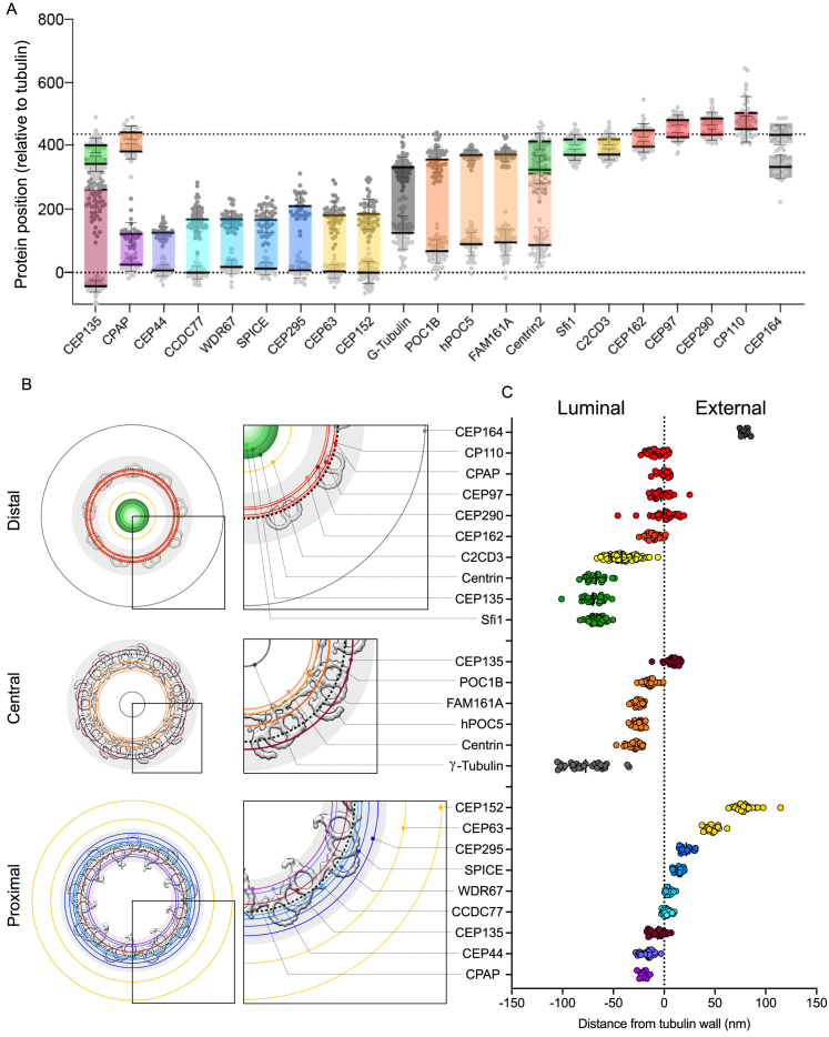
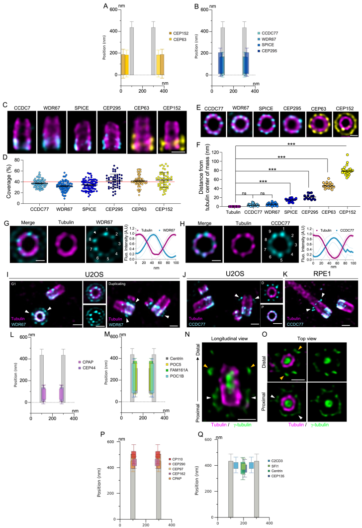
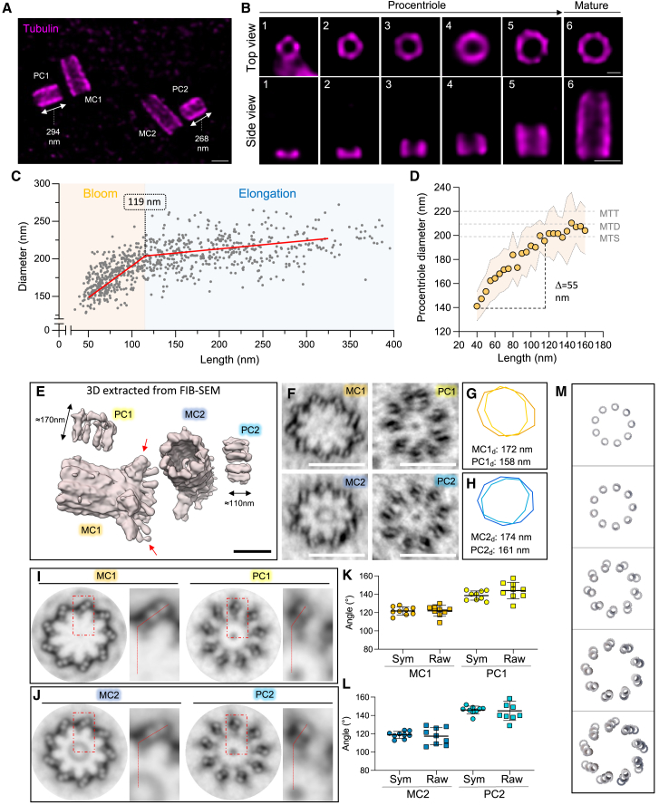
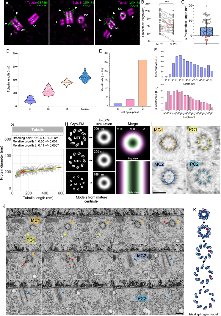
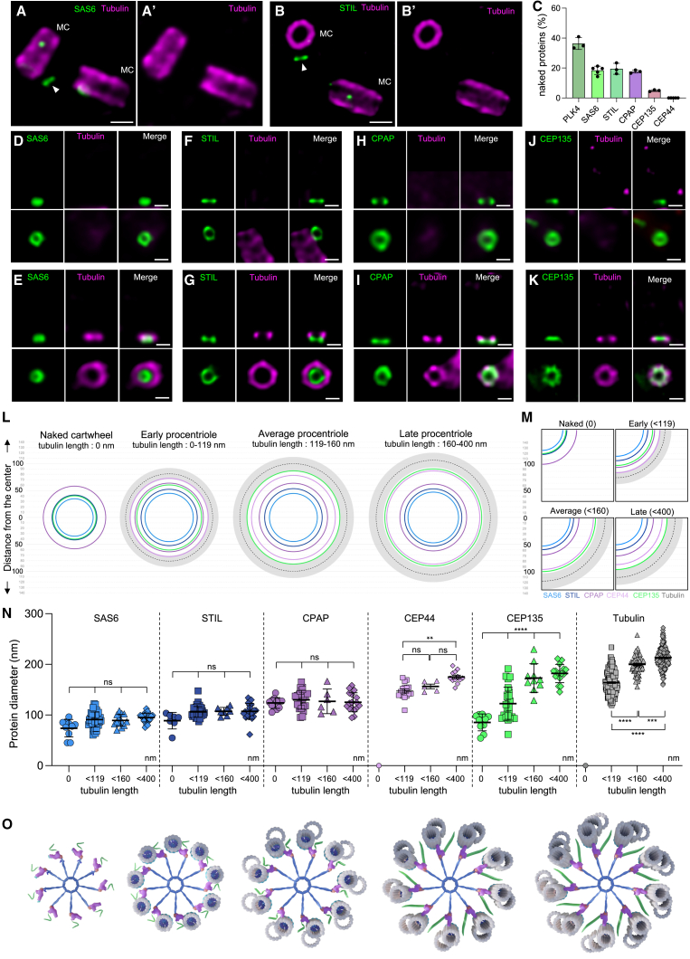
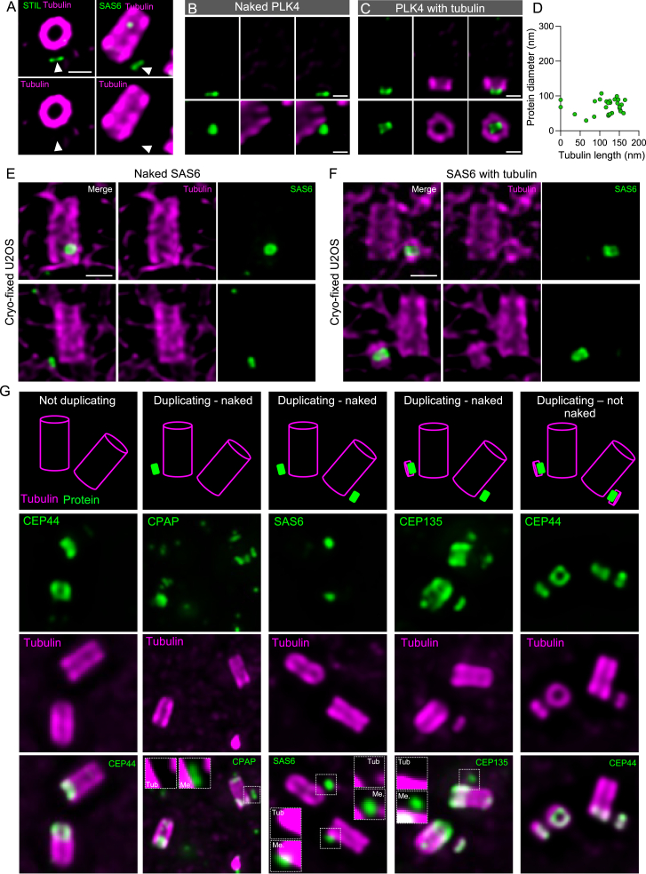
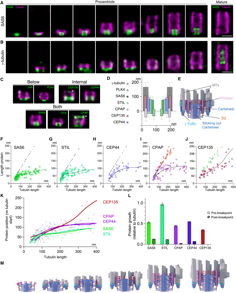
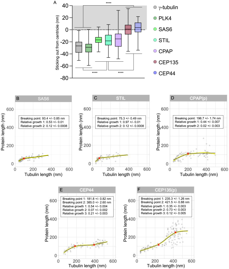
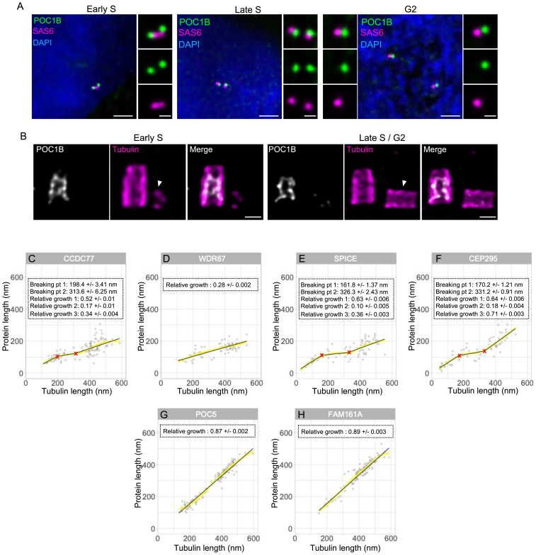
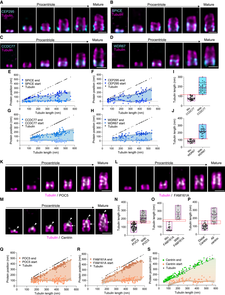
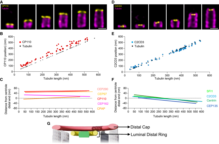
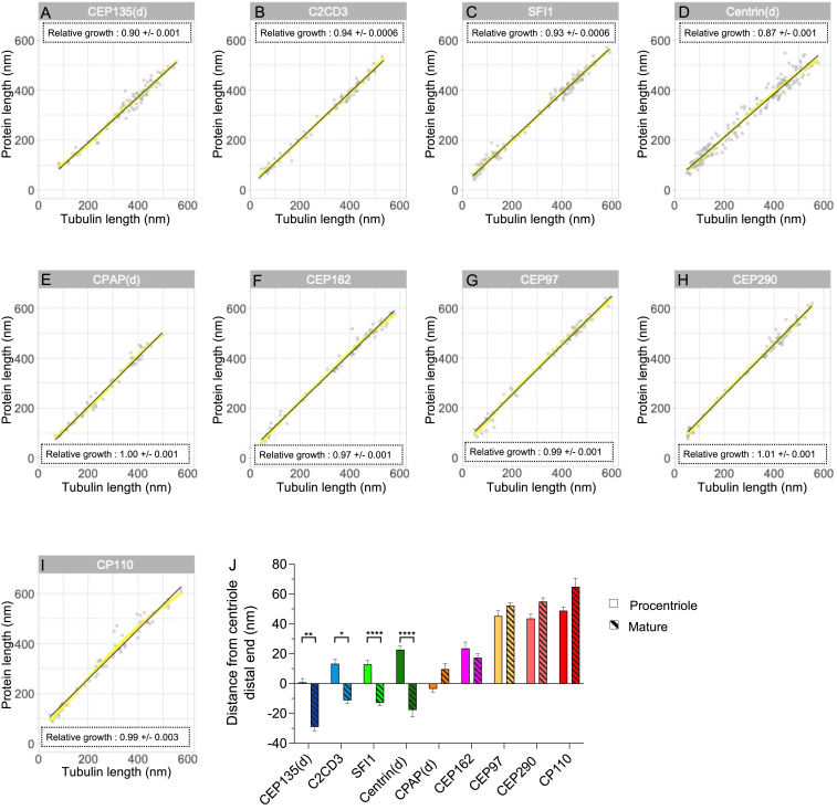
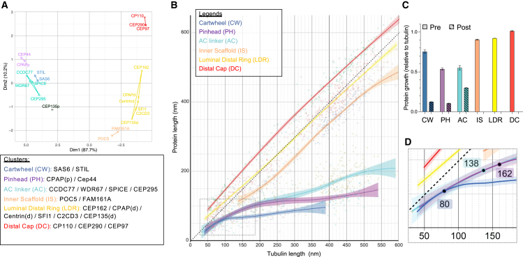
Centriole biogenesis, as in most organelle assemblies, involves the sequential recruitment of sub-structural elements that will support its function. To uncover this process, we correlated the spatial location of 24 centriolar proteins with structural features using expansion microscopy. A time-series reconstruction of protein distributions throughout human procentriole assembly unveiled the molecular architecture of the centriole biogenesis steps. We found that the process initiates with the formation of a naked cartwheel devoid of microtubules. Next, the bloom phase progresses with microtubule blade assembly, concomitantly with radial separation and rapid cartwheel growth. In the subsequent elongation phase, the tubulin backbone grows linearly with the recruitment of the A-C linker, followed by proteins of the inner scaffold (IS). By following six structural modules, we modeled 4D assembly of the human centriole. Collectively, this work provides a framework to investigate the spatial and temporal assembly of large macromolecules.

摘要

中心体生物发生,与大多数细胞器组装一样,涉及到支持其功能的亚结构元件的顺序募集。为了揭示这一过程,我们使用扩展显微镜将 24 种中心体蛋白的空间位置与结构特征相关联。对人起始中心体组装过程中蛋白质分布的时间序列重建揭示了中心体生物发生步骤的分子结构。我们发现,该过程首先是在没有微管的情况下形成裸露的轮辐。接下来,随着微管叶片组装, bloom 阶段进展,同时伴随着径向分离和快速轮辐生长。在随后的伸长阶段,微管骨架线性生长,同时招募 A-C 接头,随后是内支架(IS)的蛋白质。通过跟踪六个结构模块,我们对人中心体的 4D 组装进行了建模。总的来说,这项工作为研究大型大分子的空间和时间组装提供了一个框架。

https://cdn.ncbi.nlm.nih.gov/pmc/blobs/5d5b/11060037/6da8ab04a17c/figs9.jpg
https://cdn.ncbi.nlm.nih.gov/pmc/blobs/5d5b/11060037/9b61a4692876/fx1.jpg
https://cdn.ncbi.nlm.nih.gov/pmc/blobs/5d5b/11060037/9dda175b4fb9/figs1.jpg
https://cdn.ncbi.nlm.nih.gov/pmc/blobs/5d5b/11060037/03d9a909e93b/gr1.jpg
https://cdn.ncbi.nlm.nih.gov/pmc/blobs/5d5b/11060037/2cc5080be1b8/figs2.jpg
https://cdn.ncbi.nlm.nih.gov/pmc/blobs/5d5b/11060037/78057ce44e9a/figs3.jpg
https://cdn.ncbi.nlm.nih.gov/pmc/blobs/5d5b/11060037/5dbbe37c9d4f/gr2.jpg
https://cdn.ncbi.nlm.nih.gov/pmc/blobs/5d5b/11060037/e994355a5541/figs4.jpg
https://cdn.ncbi.nlm.nih.gov/pmc/blobs/5d5b/11060037/bb9b35aecb12/gr3.jpg
https://cdn.ncbi.nlm.nih.gov/pmc/blobs/5d5b/11060037/95b19a900bed/figs5.jpg
https://cdn.ncbi.nlm.nih.gov/pmc/blobs/5d5b/11060037/09277fd66892/gr4.jpg
https://cdn.ncbi.nlm.nih.gov/pmc/blobs/5d5b/11060037/8990cc8c8a01/figs6.jpg
https://cdn.ncbi.nlm.nih.gov/pmc/blobs/5d5b/11060037/88c87ceb978e/figs7.jpg
https://cdn.ncbi.nlm.nih.gov/pmc/blobs/5d5b/11060037/00be7a3d27ba/gr5.jpg
https://cdn.ncbi.nlm.nih.gov/pmc/blobs/5d5b/11060037/d9cdfd26eafe/gr6.jpg
https://cdn.ncbi.nlm.nih.gov/pmc/blobs/5d5b/11060037/6f6917fc47b3/figs8.jpg
https://cdn.ncbi.nlm.nih.gov/pmc/blobs/5d5b/11060037/e21775968cab/gr7.jpg
https://cdn.ncbi.nlm.nih.gov/pmc/blobs/5d5b/11060037/6da8ab04a17c/figs9.jpg

相似文献

1
Time-series reconstruction of the molecular architecture of human centriole assembly.人类中心体组装的分子结构的时间序列重建。
Cell. 2024 Apr 25;187(9):2158-2174.e19. doi: 10.1016/j.cell.2024.03.025. Epub 2024 Apr 10.
2
SAS-6 Association with γ-Tubulin Ring Complex Is Required for Centriole Duplication in Human Cells.SAS-6 与 γ-微管蛋白环复合物的关联对于人细胞中的中心体复制是必需的。
Curr Biol. 2020 Jun 22;30(12):2395-2403.e4. doi: 10.1016/j.cub.2020.04.036. Epub 2020 May 21.
3
A Short CEP135 Splice Isoform Controls Centriole Duplication.一种短的CEP135剪接异构体控制中心粒复制。
Curr Biol. 2015 Oct 5;25(19):2591-6. doi: 10.1016/j.cub.2015.08.039. Epub 2015 Sep 24.
4
WDR90 is a centriolar microtubule wall protein important for centriole architecture integrity.WDR90 是一种中心体微管壁蛋白,对中心体结构完整性很重要。
Elife. 2020 Sep 18;9:e57205. doi: 10.7554/eLife.57205.
5
CEP295 interacts with microtubules and is required for centriole elongation.CEP295与微管相互作用,是中心粒延长所必需的。
J Cell Sci. 2016 Jul 1;129(13):2501-13. doi: 10.1242/jcs.186338. Epub 2016 May 16.
6
Role of CAP350 in centriolar tubule stability and centriole assembly.CAP350在中心粒微管稳定性和中心粒组装中的作用。
PLoS One. 2008;3(12):e3855. doi: 10.1371/journal.pone.0003855. Epub 2008 Dec 4.
7
The ubiquitin ligase FBXW7 targets the centriolar assembly protein HsSAS-6 for degradation and thereby regulates centriole duplication.泛素连接酶 FBXW7 靶向中心体组装蛋白 HsSAS-6 进行降解,从而调节中心体复制。
J Biol Chem. 2020 Apr 3;295(14):4428-4437. doi: 10.1074/jbc.AC119.012178. Epub 2020 Feb 21.
8
PLK4-phosphorylated NEDD1 facilitates cartwheel assembly and centriole biogenesis initiations.PLK4 磷酸化的 NEDD1 促进轮辐装配和中心体发生起始。
J Cell Biol. 2021 Jan 4;220(1). doi: 10.1083/jcb.202002151.
9
PLK4 promotes centriole duplication by phosphorylating STIL to link the procentriole cartwheel to the microtubule wall.PLK4 通过磷酸化 STIL 促进中心体复制,将前中心体车轮与微管壁连接起来。
Elife. 2019 May 22;8:e46054. doi: 10.7554/eLife.46054.
10
Centriole assembly at a glance.中心体组装速览。
J Cell Sci. 2019 Feb 20;132(4):jcs228833. doi: 10.1242/jcs.228833.

引用本文的文献

1
Non-uniform impact of extracellular osmotic variations at subcellular level.细胞外渗透压变化在亚细胞水平的非均匀影响。
Cell Death Discov. 2025 Aug 27;11(1):410. doi: 10.1038/s41420-025-02703-6.
2
Centriole Duplication at the Crossroads of Cell Cycle Control and Oncogenesis.细胞周期调控与肿瘤发生交叉点上的中心粒复制
Cells. 2025 Jul 17;14(14):1094. doi: 10.3390/cells14141094.
3
The A-C linker controls centriole structural integrity and duplication.A-C连接体控制中心粒的结构完整性和复制。

本文引用的文献

1
Poc1 bridges basal body inner junctions to promote triplet microtubule integrity and connections.Poc1 桥接基体内部连接,以促进三联微管的完整性和连接。
J Cell Biol. 2024 Aug 5;223(8). doi: 10.1083/jcb.202311104. Epub 2024 May 14.
2
Centrosomal organization of Cep152 provides flexibility in Plk4 and procentriole positioning.中心体组织的 Cep152 为 Plk4 和中心粒定位提供了灵活性。
J Cell Biol. 2023 Dec 4;222(12). doi: 10.1083/jcb.202301092. Epub 2023 Sep 14.
3
ELI trifocal microscope: a precise system to prepare target cryo-lamellae for in situ cryo-ET study.
Nat Commun. 2025 Jul 24;16(1):6836. doi: 10.1038/s41467-025-62154-6.
4
Centriole biogenesis is seeded by CEP152-CEP63-PCNT aggregates propagating outside the centriole through the Alström syndrome protein ALMS1.中心粒的发生由CEP152-CEP63-PCNT聚集体引发,这些聚集体通过阿尔斯特伦综合征蛋白ALMS1在中心粒外传播。
bioRxiv. 2025 Jun 27:2025.06.26.661604. doi: 10.1101/2025.06.26.661604.
5
The Luminal Ring Protein C2CD3 Acts as a Radial In-to-Out Organizer of the Distal Centriole and Appendages.管腔环蛋白C2CD3作为远端中心粒和附属物的径向由内向外组织者。
bioRxiv. 2025 Jun 18:2025.06.17.660204. doi: 10.1101/2025.06.17.660204.
6
In situ cryo-electron tomography reveals the progressive biogenesis of basal bodies and cilia in mouse ependymal cells.原位冷冻电子断层扫描揭示了小鼠室管膜细胞中基体和纤毛的渐进生物发生过程。
Nat Commun. 2025 Jul 1;16(1):5932. doi: 10.1038/s41467-025-61015-6.
7
Axonemal Dynein Visualized in Primary Cilia via Expansion Microscopy.通过扩展显微镜技术在初级纤毛中可视化轴丝动力蛋白。
Cytoskeleton (Hoboken). 2025 Jun 24. doi: 10.1002/cm.70004.
8
C. elegans SSNA-1 is required for the structural integrity of centrioles and bipolar spindle assembly.秀丽隐杆线虫的SSNA-1是中心粒的结构完整性和双极纺锤体组装所必需的。
Nat Commun. 2025 Jun 5;16(1):5220. doi: 10.1038/s41467-025-59939-0.
9
TRIM37 prevents ectopic spindle pole assembly by peptide motif recognition and substrate-dependent oligomerization.TRIM37通过肽基序识别和底物依赖性寡聚化来防止异位纺锤体极组装。
Nat Struct Mol Biol. 2025 May 25. doi: 10.1038/s41594-025-01562-0.
10
Synchronized temporal-spatial analysis via microscopy and phosphoproteomics (STAMP) of quiescence.通过显微镜和磷酸化蛋白质组学对静止状态进行同步时空分析(STAMP)。
Sci Adv. 2025 Apr 25;11(17):eadt9712. doi: 10.1126/sciadv.adt9712.
ELI 三焦点显微镜:用于原位冷冻电镜研究的目标冷冻薄片制备的精确系统。
Nat Methods. 2023 Feb;20(2):276-283. doi: 10.1038/s41592-022-01748-0. Epub 2023 Jan 16.
4
Integrated multimodality microscope for accurate and efficient target-guided cryo-lamellae preparation.用于精确且高效的靶向冷冻薄片制备的集成多模态显微镜。
Nat Methods. 2023 Feb;20(2):268-275. doi: 10.1038/s41592-022-01749-z. Epub 2023 Jan 16.
5
Human SFI1 and Centrin form a complex critical for centriole architecture and ciliogenesis.人 SFI1 和 Centrin 形成了一个对中心体结构和纤毛发生至关重要的复合物。
EMBO J. 2022 Nov 2;41(21):e112107. doi: 10.15252/embj.2022112107. Epub 2022 Sep 20.
6
CPAP insufficiency leads to incomplete centrioles that duplicate but fragment.CPAP 不足导致不完全中心体复制但片段化。
J Cell Biol. 2022 May 2;221(5). doi: 10.1083/jcb.202108018. Epub 2022 Apr 9.
7
Visualizing the native cellular organization by coupling cryofixation with expansion microscopy (Cryo-ExM).通过将冷冻固定与扩展显微镜技术(Cryo-ExM)相结合来可视化原生细胞组织。
Nat Methods. 2022 Feb;19(2):216-222. doi: 10.1038/s41592-021-01356-4. Epub 2022 Jan 13.
8
Sub-centrosomal mapping identifies augmin-γTuRC as part of a centriole-stabilizing scaffold.亚中心体定位将 augmin-γTuRC 鉴定为中心粒稳定支架的一部分。
Nat Commun. 2021 Oct 15;12(1):6042. doi: 10.1038/s41467-021-26252-5.
9
An open-access volume electron microscopy atlas of whole cells and tissues.开放获取的全细胞和组织电子显微镜图谱集
Nature. 2021 Nov;599(7883):147-151. doi: 10.1038/s41586-021-03992-4. Epub 2021 Oct 6.
10
Ultrastructure expansion microscopy (U-ExM).超微结构扩展显微镜(U-ExM)。
Methods Cell Biol. 2021;161:57-81. doi: 10.1016/bs.mcb.2020.05.006. Epub 2020 Jul 1.