• 文献检索
  • 文档翻译
  • 深度研究
  • 学术资讯
  • Suppr Zotero 插件Zotero 插件
  • 邀请有礼
  • 套餐&价格
  • 历史记录
应用&插件
Suppr Zotero 插件Zotero 插件浏览器插件Mac 客户端Windows 客户端微信小程序
定价
高级版会员购买积分包购买API积分包
服务
文献检索文档翻译深度研究API 文档MCP 服务
关于我们
关于 Suppr公司介绍联系我们用户协议隐私条款
关注我们

Suppr 超能文献

核心技术专利:CN118964589B侵权必究
粤ICP备2023148730 号-1Suppr @ 2026

文献检索

告别复杂PubMed语法,用中文像聊天一样搜索,搜遍4000万医学文献。AI智能推荐,让科研检索更轻松。

立即免费搜索

文件翻译

保留排版,准确专业,支持PDF/Word/PPT等文件格式,支持 12+语言互译。

免费翻译文档

深度研究

AI帮你快速写综述,25分钟生成高质量综述,智能提取关键信息,辅助科研写作。

立即免费体验

基于微阵列的结肠癌腹膜转移动物模型耐药性检测及表达分析。

Microarray-based detection and expression analysis of drug resistance in an animal model of peritoneal metastasis from colon cancer.

机构信息

Department of Surgery, Medical Faculty Mannheim, Universitätsmedizin Mannheim, Heidelberg University, Theodor-Kutzer-Ufer 1-3, 68167, Mannheim, Germany.

Laboratory of Human Genetics, Genetic Resources Institute of Ministry of Science and Education, Baku, Azerbaijan.

出版信息

Clin Exp Metastasis. 2024 Oct;41(5):707-715. doi: 10.1007/s10585-024-10283-5. Epub 2024 Apr 12.

DOI:10.1007/s10585-024-10283-5
PMID:38609535
原文链接:https://pmc.ncbi.nlm.nih.gov/articles/PMC11499332/
Abstract

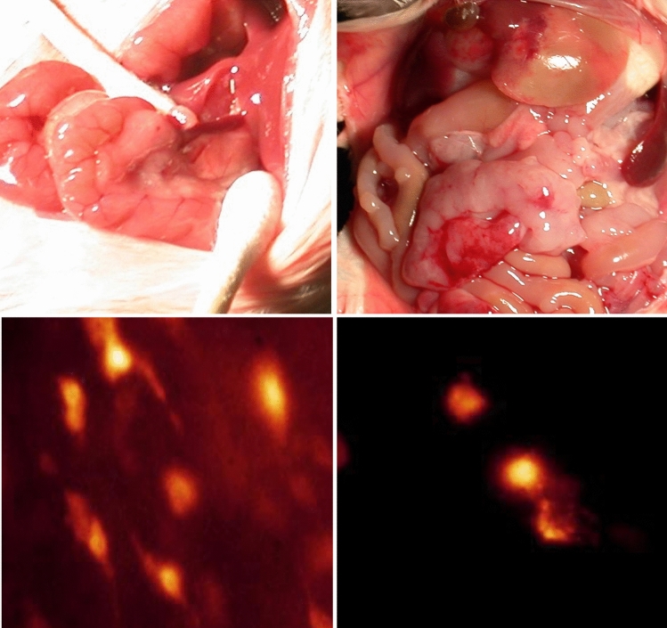
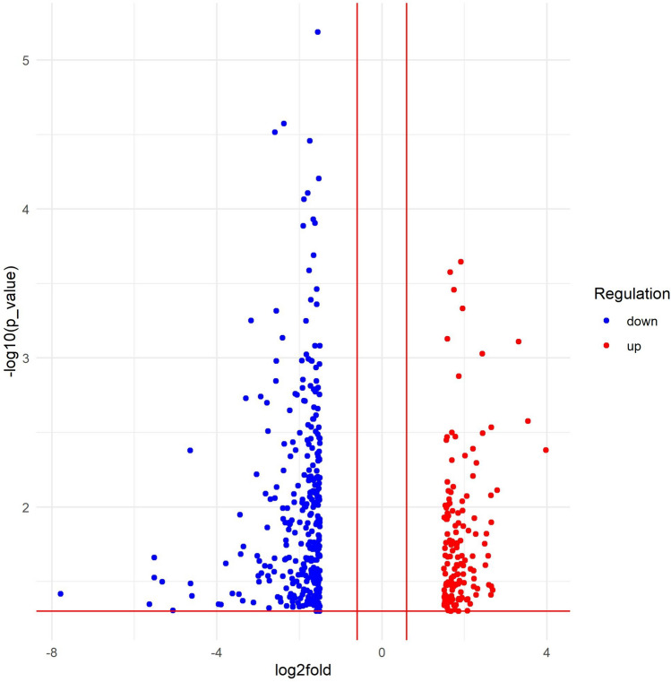
Chemotherapy drugs efficiently eradicate rapidly dividing differentiated cells by inducing cell death, but poorly target slowly dividing cells, including cancer stem cells and dormant cancer cells, in the later course of treatment. Prolonged exposure to chemotherapy results in a decrease in the proportion of apoptotic cells in the tumour mass. To investigate and characterize the molecular basis of this phenomenon, microarray-based expression analysis was performed to compare tHcred-DEVD-EGFP-caspase 3-sensor transfected C-26 tumour cells that were harvested after engraftment into mice treated with or without 5-FU. Peritoneal metastasis was induced by intraperitoneal injection of C-26 cells, which were subsequently reisolated from omental metastatic tumours after the mice were sacrificed by the end of the 10th day after tumour injection. The purity of reisolated tHcred2-DEVD-EGFP-caspase 3-sensor-expressing C-26 cells was confirmed using FLIM, and total RNA was extracted for gene expression profiling. The validation of relative transcript levels was carried out via real-time semiquantitative RT‒PCR assays. Our results demonstrated that chemotherapy induced the differential expression of mediators of cancer cell dormancy and cell survival-related genes and downregulation of both intrinsic and extrinsic apoptotic signalling pathways. Despite the fact that some differentially expressed genes, such as BMP7 and Prss11, have not been thoroughly studied in the context of chemoresistance thus far, they might be potential candidates for future studies on overcoming drug resistance.

摘要

化疗药物通过诱导细胞死亡有效地消灭快速分裂的分化细胞,但在治疗后期对缓慢分裂的细胞(包括癌症干细胞和休眠癌细胞)的靶向作用较差。长期暴露于化疗会导致肿瘤组织中凋亡细胞的比例下降。为了研究和描述这种现象的分子基础,我们进行了基于微阵列的表达分析,比较了在接受或不接受 5-FU 治疗的小鼠中植入的 tHcred-DEVD-EGFP-caspase 3-传感器转染的 C-26 肿瘤细胞。通过腹腔内注射 C-26 细胞诱导腹膜转移,然后在肿瘤注射后第 10 天处死小鼠时,从网膜转移性肿瘤中重新分离这些细胞。使用 FLIM 确认重新分离的 tHcred2-DEVD-EGFP-caspase 3-传感器表达的 C-26 细胞的纯度,并提取总 RNA 进行基因表达谱分析。通过实时半定量 RT-PCR 测定验证相对转录水平。我们的结果表明,化疗诱导了癌症细胞休眠和细胞存活相关基因的介质的差异表达,并下调了内在和外在凋亡信号通路。尽管一些差异表达的基因,如 BMP7 和 Prss11,迄今为止在化疗耐药性方面尚未得到深入研究,但它们可能是未来克服药物耐药性研究的潜在候选基因。

https://cdn.ncbi.nlm.nih.gov/pmc/blobs/225f/11499332/e0664c7c7ddf/10585_2024_10283_Fig2_HTML.jpg
https://cdn.ncbi.nlm.nih.gov/pmc/blobs/225f/11499332/ec16c9f7a619/10585_2024_10283_Fig1_HTML.jpg
https://cdn.ncbi.nlm.nih.gov/pmc/blobs/225f/11499332/e0664c7c7ddf/10585_2024_10283_Fig2_HTML.jpg
https://cdn.ncbi.nlm.nih.gov/pmc/blobs/225f/11499332/ec16c9f7a619/10585_2024_10283_Fig1_HTML.jpg
https://cdn.ncbi.nlm.nih.gov/pmc/blobs/225f/11499332/e0664c7c7ddf/10585_2024_10283_Fig2_HTML.jpg

相似文献

1
Microarray-based detection and expression analysis of drug resistance in an animal model of peritoneal metastasis from colon cancer.基于微阵列的结肠癌腹膜转移动物模型耐药性检测及表达分析。
Clin Exp Metastasis. 2024 Oct;41(5):707-715. doi: 10.1007/s10585-024-10283-5. Epub 2024 Apr 12.
2
Downregulation of lncRNA CCAT1 enhances 5-fluorouracil sensitivity in human colon cancer cells.长链非编码 RNA CCAT1 的下调增强了人结肠癌细胞对 5-氟尿嘧啶的敏感性。
BMC Mol Cell Biol. 2019 Apr 23;20(1):9. doi: 10.1186/s12860-019-0188-1.
3
INHBA regulates Hippo signaling to confer 5-FU chemoresistance mediated by cellular senescence in colon cancer cells.INHBA 通过细胞衰老调控 Hippo 信号通路赋予结直肠癌细胞对 5-FU 的化疗耐药性。
Int J Biochem Cell Biol. 2024 Jun;171:106570. doi: 10.1016/j.biocel.2024.106570. Epub 2024 Apr 7.
4
Comparison of gene expression in HCT116 treatment derivatives generated by two different 5-fluorouracil exposure protocols.两种不同5-氟尿嘧啶暴露方案产生的HCT116处理衍生物中基因表达的比较。
Mol Cancer. 2004 Apr 26;3:11. doi: 10.1186/1476-4598-3-11.
5
Knockdown of PRDX2 sensitizes colon cancer cells to 5-FU by suppressing the PI3K/AKT signaling pathway.敲低PRDX2通过抑制PI3K/AKT信号通路使结肠癌细胞对5-氟尿嘧啶敏感。
Biosci Rep. 2017 May 11;37(3). doi: 10.1042/BSR20160447. Print 2017 Jun 30.
6
The combination of FLCWK with 5-FU inhibits colon cancer and multidrug resistance by activating PXR to suppress the IL-6/STAT3 pathway.FLCWK 与 5-FU 的联合应用通过激活 PXR 抑制 IL-6/STAT3 通路来抑制结肠癌和多药耐药。
J Cell Mol Med. 2024 Nov;28(21):e70185. doi: 10.1111/jcmm.70185.
7
MicroRNA-23a antisense enhances 5-fluorouracil chemosensitivity through APAF-1/caspase-9 apoptotic pathway in colorectal cancer cells.miRNA-23a 反义寡核苷酸通过 APAF-1/caspase-9 凋亡途径增强结直肠癌细胞对 5-氟尿嘧啶的化疗敏感性。
J Cell Biochem. 2014 Apr;115(4):772-84. doi: 10.1002/jcb.24721.
8
The c-MYC-ABCB5 axis plays a pivotal role in 5-fluorouracil resistance in human colon cancer cells.c-MYC-ABCB5轴在人结肠癌细胞对5-氟尿嘧啶的耐药性中起关键作用。
J Cell Mol Med. 2015 Jul;19(7):1569-81. doi: 10.1111/jcmm.12531. Epub 2015 Feb 17.
9
Curcumin synergizes with 5-fluorouracil by impairing AMPK/ULK1-dependent autophagy, AKT activity and enhancing apoptosis in colon cancer cells with tumor growth inhibition in xenograft mice.姜黄素通过损害 AMPK/ULK1 依赖性自噬、AKT 活性和增强结肠癌细胞中的细胞凋亡,与 5-氟尿嘧啶协同作用,并抑制异种移植小鼠肿瘤生长。
J Exp Clin Cancer Res. 2017 Dec 22;36(1):190. doi: 10.1186/s13046-017-0661-7.
10
MicroRNA-494 sensitizes colon cancer cells to fluorouracil through regulation of DPYD.微小RNA-494通过调控二氢嘧啶脱氢酶使结肠癌细胞对氟尿嘧啶敏感。
IUBMB Life. 2015 Mar;67(3):191-201. doi: 10.1002/iub.1361. Epub 2015 Apr 15.

本文引用的文献

1
The present and future of PI3K inhibitors for cancer therapy.用于癌症治疗的PI3K抑制剂的现状与未来。
Nat Cancer. 2021 Jun;2(6):587-597. doi: 10.1038/s43018-021-00218-4. Epub 2021 Jun 17.
2
Drug resistance and combating drug resistance in cancer.癌症中的耐药性与抗耐药性
Cancer Drug Resist. 2019;2(2):141-160. doi: 10.20517/cdr.2019.10. Epub 2019 Jun 19.
3
The MAPK and AMPK signalings: interplay and implication in targeted cancer therapy.MAPK 和 AMPK 信号通路:在肿瘤靶向治疗中的相互作用和影响。
J Hematol Oncol. 2020 Aug 17;13(1):113. doi: 10.1186/s13045-020-00949-4.
4
Targeting chemoresistant colorectal cancer via systemic administration of a BMP7 variant.通过系统给予 BMP7 变体靶向化疗耐药结直肠癌。
Oncogene. 2020 Jan;39(5):987-1003. doi: 10.1038/s41388-019-1047-4. Epub 2019 Oct 7.
5
Targeting PI3K in cancer: mechanisms and advances in clinical trials.针对癌症的 PI3K 靶向治疗:机制与临床试验进展。
Mol Cancer. 2019 Feb 19;18(1):26. doi: 10.1186/s12943-019-0954-x.
6
Dormancy and cancer stem cells: An enigma for cancer therapeutic targeting.休眠和癌症干细胞:癌症治疗靶向的谜团。
Adv Cancer Res. 2019;141:43-84. doi: 10.1016/bs.acr.2018.12.002. Epub 2019 Jan 16.
7
Targeting Cancer Cell Dormancy.靶向肿瘤休眠细胞
Trends Pharmacol Sci. 2019 Feb;40(2):128-141. doi: 10.1016/j.tips.2018.12.004. Epub 2019 Jan 3.
8
Epigenetically upregulated oncoprotein PLCE1 drives esophageal carcinoma angiogenesis and proliferation via activating the PI-PLCε-NF-κB signaling pathway and VEGF-C/ Bcl-2 expression.表观上调的癌蛋白 PLCE1 通过激活 PI-PLCε-NF-κB 信号通路和 VEGF-C/Bcl-2 表达,驱动食管癌血管生成和增殖。
Mol Cancer. 2019 Jan 4;18(1):1. doi: 10.1186/s12943-018-0930-x.
9
PI3K/Akt signaling transduction pathway, erythropoiesis and glycolysis in hypoxia (Review).PI3K/Akt 信号转导通路、低氧状态下的红细胞生成和糖酵解(综述)。
Mol Med Rep. 2019 Feb;19(2):783-791. doi: 10.3892/mmr.2018.9713. Epub 2018 Dec 3.
10
Global cancer statistics 2018: GLOBOCAN estimates of incidence and mortality worldwide for 36 cancers in 185 countries.全球癌症统计数据 2018:GLOBOCAN 对全球 185 个国家/地区 36 种癌症的发病率和死亡率的估计。
CA Cancer J Clin. 2018 Nov;68(6):394-424. doi: 10.3322/caac.21492. Epub 2018 Sep 12.