Department of Pathology and Key Laboratory for Xinjiang Endemic and Ethnic Diseases, The First Affiliated Hospital, Shihezi University School of Medicine, Shihezi, 832002, China.
The people's hospital of Suzhou National Hi-Tech District, Suzhou, 215010, China.
Mol Cancer. 2019 Jan 4;18(1):1. doi: 10.1186/s12943-018-0930-x.
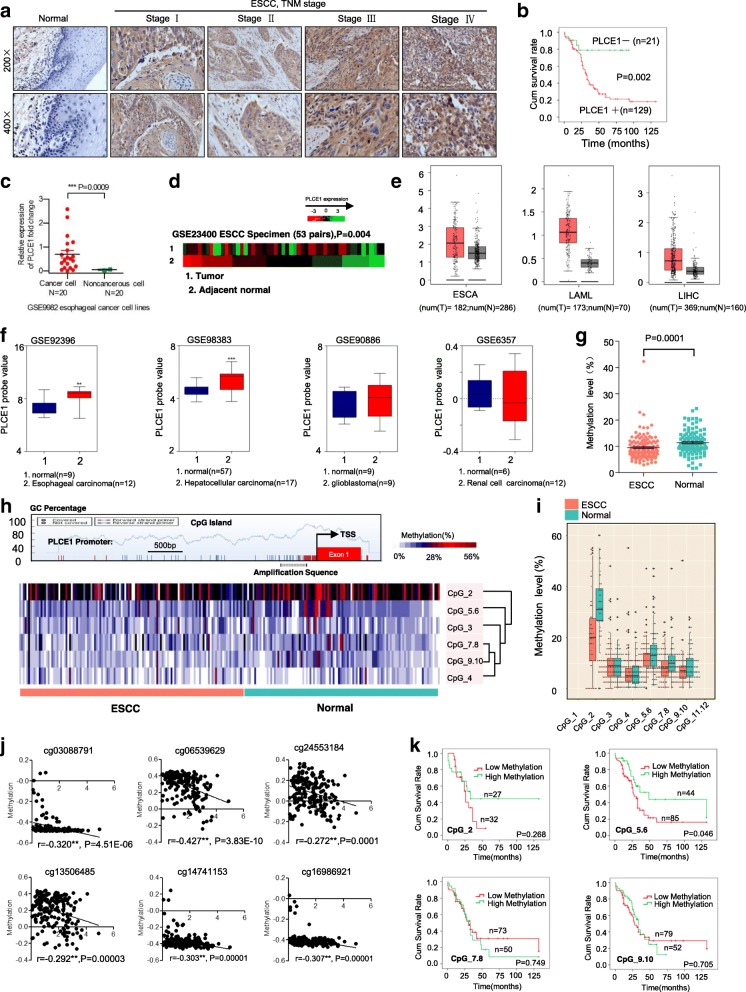
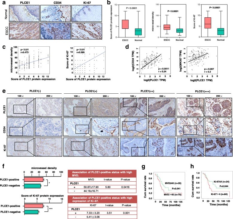
Esophageal squamous cell carcinoma (ESCC) is one of the most lethal malignancies. Neovascularization during tumorigenesis supplies oxygen and nutrients to proliferative tumor cells, and serves as a conduit for migration. Targeting oncogenes involved in angiogenesis is needed to treat organ-confined and locally advanced ESCC. Although the phospholipase C epsilon-1 (PLCE1) gene was originally identified as a susceptibility gene for ESCC, how PLCE1 is involved in ESCC is unclear.
Matrix-assisted laser desorption ionization time-of-flight mass spectrometry were used to measure the methylation status of the PLCE1 promoter region. To validate the underlying mechanism for PLCE1 in constitutive activation of the NF-κB signaling pathway, we performed studies using in vitro and in vivo assays and samples from 368 formalin-fixed esophageal cancer tissues and 215 normal tissues with IHC using tissue microarrays and the Cancer Genome Atlas dataset.
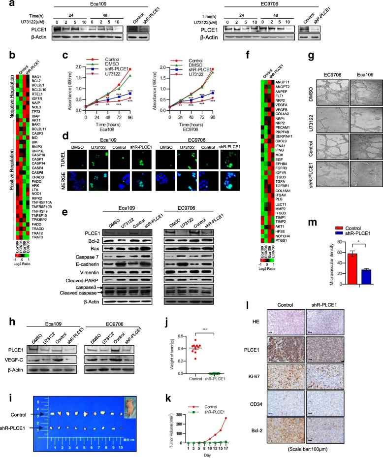
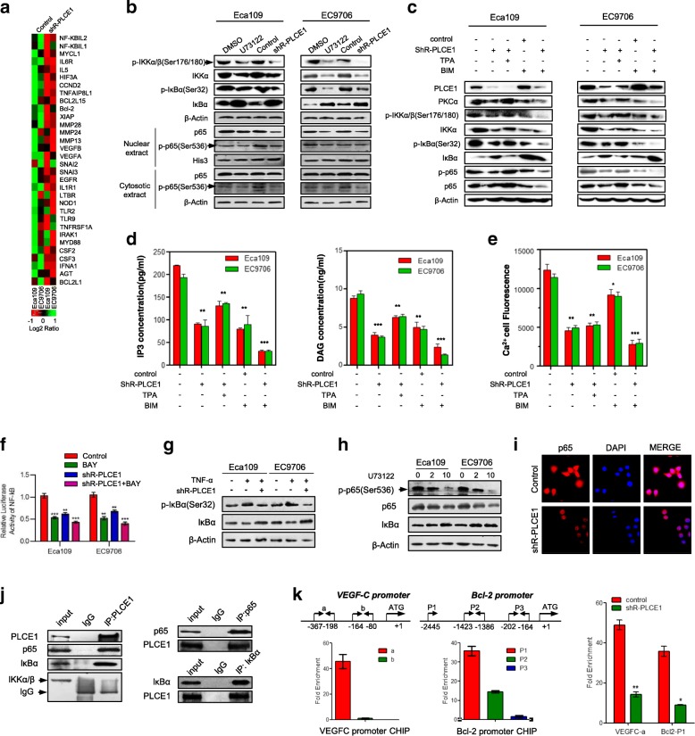
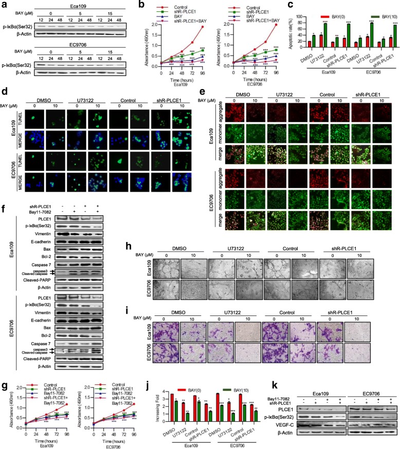
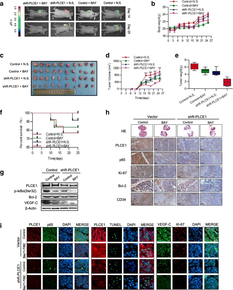
We report that hypomethylation-associated up-regulation of PLCE1 expression was correlated with tumor angiogenesis and poor prognosis in ESCC cohorts. PLCE1 can activate NF-κB through phosphoinositide-phospholipase C-ε (PI-PLCε) signaling pathway. Furthermore, PLCE1 can bind p65 and IκBα proteins, promoting IκBα-S32 and p65-S536 phosphorylation. Consequently, phosphorylated IκBα promotes nuclear translocation of p50/p65 and p65, as a transcription factor, can bind vascular endothelial growth factor-C and bcl-2 promoters, enhancing angiogenesis and inhibiting apoptosis in vitro. Moreover, xenograft tumors in nude mice proved that PLCE1 can induce angiogenesis, inhibit apoptosis, and increase tumor aggressiveness via the NF-κB signaling pathway in vivo.
Our findings not only provide evidence that hypomethylation-induced PLCE1 confers angiogenesis and proliferation in ESCC by activating PI-PLCε-NF-κB signaling pathway and VEGF-C/Bcl-2 expression, but also suggest that modulation of PLCE1 by epigenetic modification or a selective inhibitor may be a promising therapeutic approach for the treatment of ESCC.
食管鳞状细胞癌(ESCC)是最致命的恶性肿瘤之一。肿瘤发生过程中的新生血管为增殖性肿瘤细胞提供氧气和营养,并作为迁移的通道。针对参与血管生成的癌基因是治疗局限性和局部进展性 ESCC 的必要手段。虽然磷脂酶 C epsilon-1(PLCE1)基因最初被确定为 ESCC 的易感性基因,但 PLCE1 如何参与 ESCC 尚不清楚。
基质辅助激光解吸电离飞行时间质谱法测量 PLCE1 启动子区域的甲基化状态。为了验证 PLCE1 在 NF-κB 信号通路组成性激活中的潜在机制,我们使用体外和体内实验以及组织微阵列和癌症基因组图谱数据集的 368 例福尔马林固定食管癌细胞组织和 215 例正常组织的免疫组织化学研究进行了研究。
我们报告说,PLCE1 表达的低甲基化相关上调与 ESCC 队列中的肿瘤血管生成和不良预后相关。PLCE1 可以通过磷酸肌醇磷脂酶 C-ε(PI-PLCε)信号通路激活 NF-κB。此外,PLCE1 可以与 p65 和 IκBα 蛋白结合,促进 IκBα-S32 和 p65-S536 磷酸化。因此,磷酸化的 IκBα 促进 p50/p65 的核转位,作为转录因子的 p65 可以结合血管内皮生长因子-C 和 bcl-2 启动子,增强体外的血管生成和抑制凋亡。此外,裸鼠异种移植肿瘤证明,PLCE1 可以通过 NF-κB 信号通路在体内诱导血管生成、抑制凋亡和增加肿瘤侵袭性。
我们的研究结果不仅提供了证据表明,低甲基化诱导的 PLCE1 通过激活 PI-PLCε-NF-κB 信号通路和 VEGF-C/Bcl-2 表达,在 ESCC 中赋予血管生成和增殖能力,还表明通过表观遗传修饰或选择性抑制剂调节 PLCE1 可能是治疗 ESCC 的一种有前途的治疗方法。