Center for Cognition and Brain Disorders, The Affiliated Hospital of Hangzhou Normal University, No. 2318, Yuhangtang Rd, Hangzhou, 311121, Zhejiang Province, People's Republic of China.
Institute of Psychological Science, Hangzhou Normal University, Hangzhou, Zhejiang Province, People's Republic of China.
Sci Rep. 2024 Apr 18;14(1):8940. doi: 10.1038/s41598-024-56866-w.
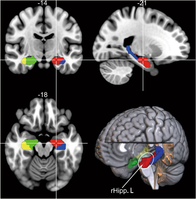
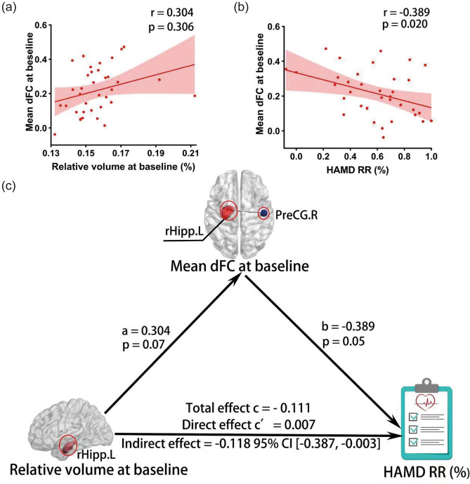
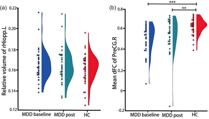
An abnormality of structures and functions in the hippocampus may have a key role in the pathophysiology of major depressive disorder (MDD). However, it is unclear whether structure factors of the hippocampus effectively impact antidepressant responses by hippocampal functional activity in MDD patients. We collected longitudinal data from 36 MDD patients before and after a 3-month course of antidepressant pharmacotherapy. Additionally, we obtained baseline data from 43 healthy controls matched for sex and age. Using resting-state functional magnetic resonance imaging (rs-fMRI), we estimated the dynamic functional connectivity (dFC) of the hippocampal subregions using a sliding-window method. The gray matter volume was calculated using voxel-based morphometry (VBM). The results indicated that patients with MDD exhibited significantly lower dFC of the left rostral hippocampus (rHipp.L) with the right precentral gyrus, left superior temporal gyrus and left postcentral gyrus compared to healthy controls at baseline. In MDD patients, the dFC of the rHipp.L with right precentral gyrus at baseline was correlated with both the rHipp.L volume and HAMD remission rate, and also mediated the effects of the rHipp.L volume on antidepressant performance. Our findings suggested that the interaction between hippocampal structure and functional activity might affect antidepressant performance, which provided a novel insight into the hippocampus-related neurobiological mechanism of MDD.
海马体结构和功能的异常可能在重度抑郁症(MDD)的病理生理学中起关键作用。然而,目前尚不清楚 MDD 患者的海马体结构因素是否通过海马体功能活动有效影响抗抑郁反应。我们从 36 名接受为期 3 个月抗抑郁药物治疗的 MDD 患者中收集了纵向数据,并从 43 名性别和年龄匹配的健康对照者中获得了基线数据。使用静息态功能磁共振成像(rs-fMRI),我们使用滑动窗口方法估计了海马亚区的动态功能连接(dFC)。使用基于体素的形态测量学(VBM)计算灰质体积。结果表明,与健康对照组相比,MDD 患者在基线时左前海马体(rHipp.L)与右侧中央前回、左颞上回和左后中央回的 dFC 明显降低。在 MDD 患者中,基线时 rHipp.L 与右侧中央前回的 dFC 与 rHipp.L 体积和 HAMD 缓解率均相关,并且还介导了 rHipp.L 体积对抗抑郁作用的影响。我们的研究结果表明,海马体结构和功能活动之间的相互作用可能会影响抗抑郁反应,这为 MDD 的海马体相关神经生物学机制提供了新的见解。