Centre for Inflammatory Disease, Imperial College London, Hammersmith Hospital, Du Cane Road, London, W12 0NN, UK; Imperial College Healthcare NHS Trust, St. Mary's Hospital, Praed Street, London, W2 1NY, UK.
Centre for Computational Biology and Program in Cardiovascular and Metabolic Disorders, Duke-NUS Medical School, Singapore, Singapore.
EBioMedicine. 2024 May;103:105127. doi: 10.1016/j.ebiom.2024.105127. Epub 2024 Apr 26.
Obesity drives maladaptive changes in the white adipose tissue (WAT) which can progressively cause insulin resistance, type 2 diabetes mellitus (T2DM) and metabolic dysfunction-associated liver disease (MASLD). Obesity-mediated loss of WAT homeostasis can trigger liver steatosis through dysregulated lipid pathways such as those related to polyunsaturated fatty acid (PUFA)-derived oxylipins. However, the exact relationship between oxylipins and metabolic syndrome remains elusive and cross-tissue dynamics of oxylipins are ill-defined.
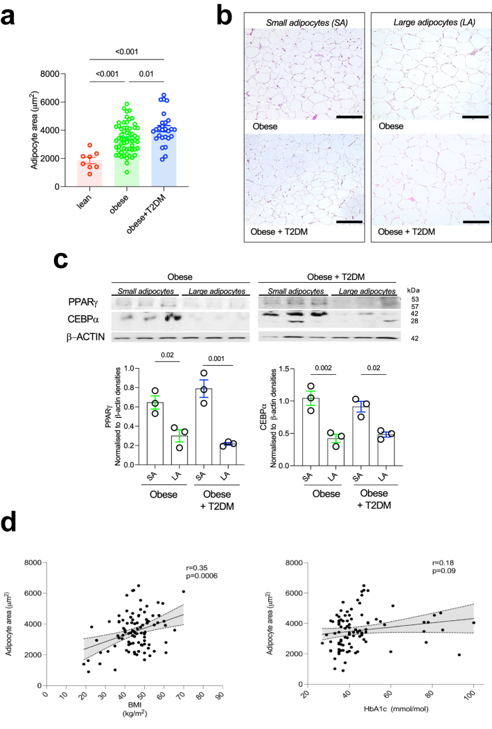
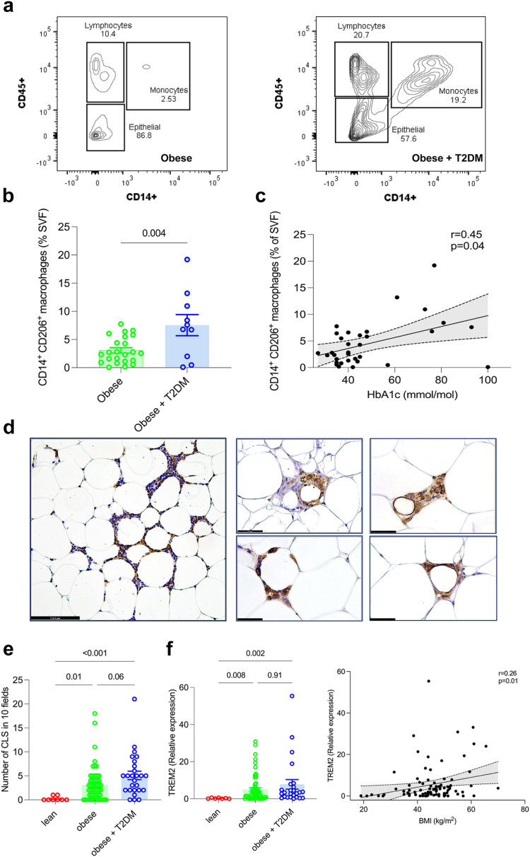
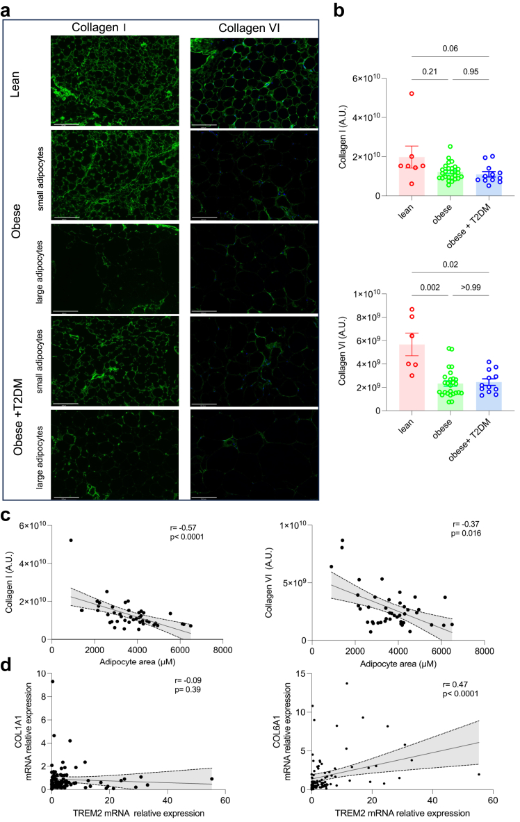
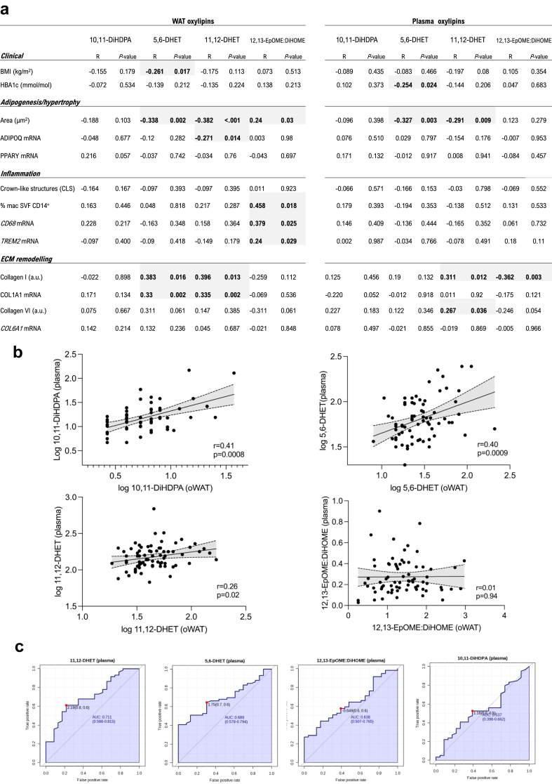
We quantified PUFA-related oxylipin species in the omental WAT, liver biopsies and plasma of 88 patients undergoing bariatric surgery (female N = 79) and 9 patients (female N = 4) undergoing upper gastrointestinal surgery, using UPLC-MS/MS. We integrated oxylipin abundance with WAT phenotypes (adipogenesis, adipocyte hypertrophy, macrophage infiltration, type I and VI collagen remodelling) and the severity of MASLD (steatosis, inflammation, fibrosis) quantified in each biopsy. The integrative analysis was subjected to (i) adjustment for known risk factors and, (ii) control for potential drug-effects through UPLC-MS/MS analysis of metformin-treated fat explants ex vivo.
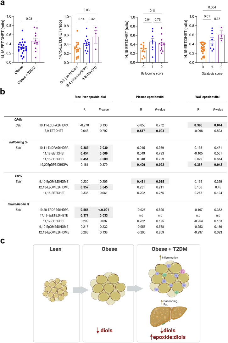
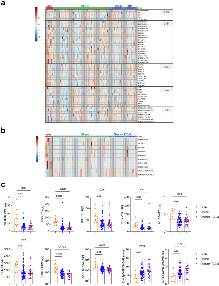
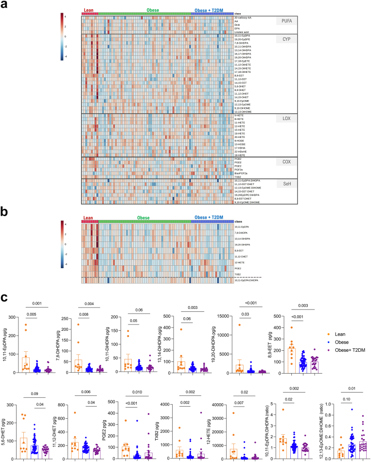
We reveal a generalized down-regulation of cytochrome P450 (CYP)-derived diols during obesity conserved between the WAT and plasma. Notably, epoxide:diol ratio, indicative of soluble epoxide hydrolyse (sEH) activity, increases with WAT inflammation/fibrosis, hepatic steatosis and T2DM. Increased 12,13-EpOME:DiHOME in WAT and liver is a marker of worsening metabolic syndrome in patients with obesity.
These findings suggest a dampened sEH activity and a possible role of fatty acid diols during metabolic syndrome in major metabolic organs such as WAT and liver. They also have implications in view of the clinical trials based on sEH inhibition for metabolic syndrome.
Wellcome Trust (PS3431_WMIH); Duke-NUS (Intramural Goh Cardiovascular Research Award (Duke-NUS-GCR/2022/0020); National Medical Research Council (OFLCG22may-0011); National Institute of Environmental Health Sciences (Z01 ES025034); NIHR Imperial Biomedical Research Centre.
肥胖会导致白色脂肪组织(WAT)发生适应性变化,从而逐渐导致胰岛素抵抗、2 型糖尿病(T2DM)和代谢功能障碍相关的肝疾病(MASLD)。肥胖介导的 WAT 稳态丧失会通过失调的脂质途径(如与多不饱和脂肪酸(PUFA)衍生的氧化脂类有关的途径)引发肝脂肪变性。然而,氧化脂类与代谢综合征的确切关系仍不清楚,并且氧化脂类在不同组织间的动态变化也不清楚。
我们使用 UPLC-MS/MS 定量分析了 88 名接受减肥手术(女性 N=79 名)和 9 名(女性 N=4 名)接受上消化道手术的患者的网膜 WAT、肝活检和血浆中的 PUFA 相关氧化脂类。我们将氧化脂类的丰度与每个活检中定量的 WAT 表型(脂肪生成、脂肪细胞肥大、巨噬细胞浸润、I 型和 VI 型胶原重塑)和 MASLD 的严重程度(脂肪变性、炎症、纤维化)整合在一起。整合分析通过(i)对已知风险因素进行调整,以及(ii)通过对体外培养的二甲双胍处理脂肪组织样本进行 UPLC-MS/MS 分析来控制潜在的药物影响。
我们发现,肥胖症期间,WAT 和血浆之间存在广泛的细胞色素 P450(CYP)衍生二醇下调。值得注意的是,环氧化物:二醇比值(指示可溶性环氧化物水解酶(sEH)活性)随着 WAT 炎症/纤维化、肝脂肪变性和 T2DM 而增加。WAT 和肝脏中 12,13-EpOME:DiHOME 的增加是肥胖患者代谢综合征恶化的标志物。
这些发现表明,在 WAT 和肝脏等主要代谢器官中,sEH 活性降低,脂肪酸二醇可能在代谢综合征中发挥作用。鉴于基于 sEH 抑制的代谢综合征临床试验,这些发现具有重要意义。
惠康信托基金会(PS3431_WMIH);杜克-新加坡国立大学(内部 Goh 心血管研究奖(Duke-NUS-GCR/2022/0020);新加坡国家医学研究理事会(OFLCG22may-0011);美国国立环境卫生科学研究所(Z01 ES025034);英国帝国理工学院生物医学研究中心(NIHR Imperial Biomedical Research Centre)。