Chiramel David Elvis, H Azeem, Santhosh Mani Emil, George Sibin, Semwal Indra, Raj R Akhila
Department of Conservative Dentistry and Endodontics, MES Dental College, Perinthalmanna, IND.
Department of Conservative Dentistry and Endodontics, Uttaranchal Dental and Medical Research Institute, Dehradun, IND.
Cureus. 2024 Mar 31;16(3):e57302. doi: 10.7759/cureus.57302. eCollection 2024 Mar.
Since the beginning of modern endodontics, there have been many concepts, strategies, and techniques for root canal preparation. A mind-boggling variety of files have developed for negotiating and shaping them throughout the years. Today's most secure, most effective, and simplest file system combines the most reliable design elements of the past with the latest technological advances to create the most effective file system. So, the need for the study is to evaluate the fracture strength of tooth roots following canal preparation by three rotary file systems: ProTaper Universal file system (Dentsply, USA), ProTaper Next file system (Dentsply Sirona USA), and Neolix A1 nickel-titanium (NiTi) file system (Orikam Healthcare India Pvt Ltd., New Delhi, India).
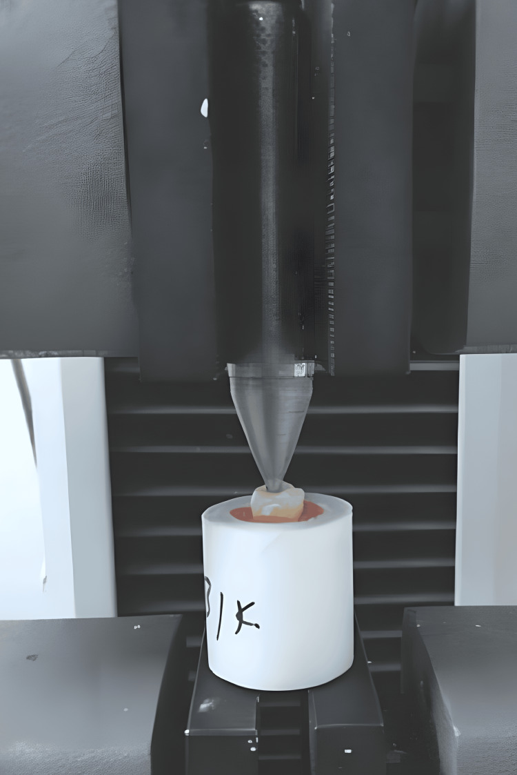
Ninety human mandibular molars were selected for the study. Inclusion criteria include human mandibular first and second molars and teeth removed for routine clinical reasons, and intact apices were selected, excluding cases with root surface caries, root surface fissures, teeth with immature root apex, mesial canal fusion, extremely short roots, thin roots, or curved roots. All teeth were preserved in a solution of 10% neutral buffered formalin for two weeks and then transferred to distilled water for examination. The teeth were randomly divided into three groups. Access cavities were created, and working lengths were determined. Groups 1, 2, and 3 underwent shaping using ProTaper Universal, ProTaper Next, and Neolix A1 (NiTi) file systems, respectively, following guidelines. Canals were irrigated with sodium hypochlorite and ethylenediaminetetraacetic acid (EDTA) and were obturated up to the mid-root region with AH Plus sealer. To facilitate fracture testing, obturation was performed to distribute the load from the spreader to the canal wall. The EndoSequence and Quick-Fill obturation system were utilized to fill the apical half of the canal with gutta-percha material. After obturation, the distal root of each tooth was cut, while the mesial root was securely positioned in a putty material. A universal testing machine was employed for the fracture tests, operating at a cross-head speed of 1 mm/min. The machine was equipped with a D11 hand spreader tip, which was inserted into the root canal to make contact with the gutta-percha. Gradual force was applied to the root canal until a fracture occurred, at which point the force application was stopped. The amount of force required to cause the fracture was measured in newtons. Data were collected and recorded using IBM SPSS Statistics for Windows, Version 17.0 (Released 2008; IBM Corp., Armonk, New York, United States) and then transferred to Microsoft Excel for analysis. Descriptive statistics, mean, and standard deviation were used for continuous data. The fracture resistance of dental roots treated with three types of files was compared using a one-way ANOVA. Graphs were generated using Excel and Word. A significance level of p<0.01 was chosen.
ANOVA indicated significant differences in mean fracture resistance: Neolix A1 (NiTi) (95.3 N) > NEXT (91.0 N) > universal (86.6 N), with a p-value of 0.004 (<0.001), confirming statistical significance.
The study concludes that the canal instrumented with Neolix A1 (NiTi) exhibits higher fracture resistance after canal instrumentation compared to ProTaper Next and ProTaper Universal.
自现代牙髓病学诞生以来,根管预备出现了许多概念、策略和技术。多年来,为了对根管进行疏通和塑形,开发出了种类繁多的锉具。如今,最安全、最有效且最简单的锉具系统是将过去最可靠的设计元素与最新技术进步相结合,从而打造出最有效的锉具系统。因此,本研究旨在评估三种旋转锉具系统对根管预备后牙根的抗折强度,这三种锉具系统分别是:ProTaper Universal锉具系统(美国登士柏公司)、ProTaper Next锉具系统(美国登士柏西诺德公司)和Neolix A1镍钛(NiTi)锉具系统(印度新德里奥里卡姆医疗保健私人有限公司)。
本研究选取了90颗人类下颌磨牙。纳入标准包括人类下颌第一和第二磨牙以及因常规临床原因拔除的牙齿,且选择根尖完整的牙齿,排除根面龋、根面裂、根尖未发育成熟、近中根管融合、牙根极短、牙根纤细或牙根弯曲的病例。所有牙齿均保存在10%中性缓冲福尔马林溶液中两周,然后转移至蒸馏水中进行检查。将牙齿随机分为三组。制备开髓洞形并确定工作长度。第1组、第2组和第3组分别按照操作指南,使用ProTaper Universal、ProTaper Next和Neolix A1(NiTi)锉具系统进行根管塑形。根管先用次氯酸钠和乙二胺四乙酸(EDTA)冲洗,然后用AH Plus封闭剂封闭至根中区域。为便于进行抗折测试,进行根管充填以使扩孔钻的负荷分散到根管壁上。采用EndoSequence和Quick-Fill充填系统用牙胶材料充填根管根尖的一半。充填后,将每颗牙齿的远中根切断,而近中根则牢固地固定在一种油灰材料中。使用万能试验机进行抗折测试,十字头速度为1 mm/min。该试验机配备了一个D11手动扩孔钻尖端,将其插入根管与牙胶接触。逐渐对根管施加力直至发生折断,此时停止施加力。记录导致折断所需的力的大小,单位为牛顿。使用IBM SPSS Statistics for Windows 17.0版(2008年发布;美国纽约州阿蒙克市IBM公司)收集和记录数据,并将其转移到Microsoft Excel中进行分析。对于连续数据,使用描述性统计、均值和标准差。使用单因素方差分析比较三种锉具处理后的牙根抗折性。使用Excel和Word生成图表。选择的显著性水平为p< 0.01。
方差分析表明平均抗折性存在显著差异:Neolix A1(NiTi)(95.3 N)> NEXT(91.0 N)> Universal(86.6 N),p值为0.004(<0.001),证实具有统计学意义。
该研究得出结论,与ProTaper Next和ProTaper Universal相比,使用Neolix A1(NiTi)进行根管预备后的根管具有更高的抗折性。