Walter and Eliza Hall Institute of Medical Research, Parkville, Melbourne, Australia.
Department of Medical Biology, University of Melbourne, Parkville, Melbourne, Australia.
PLoS Biol. 2024 May 2;22(5):e3002617. doi: 10.1371/journal.pbio.3002617. eCollection 2024 May.
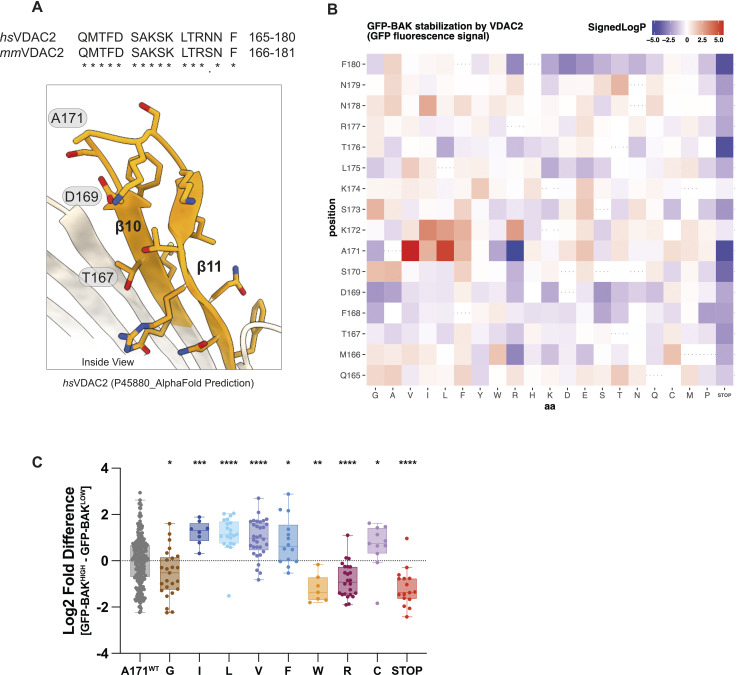
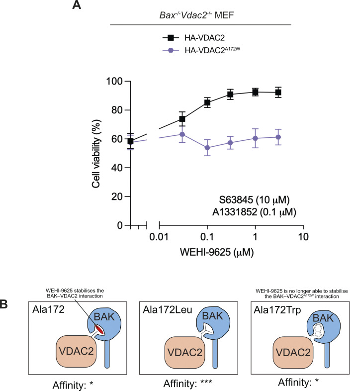
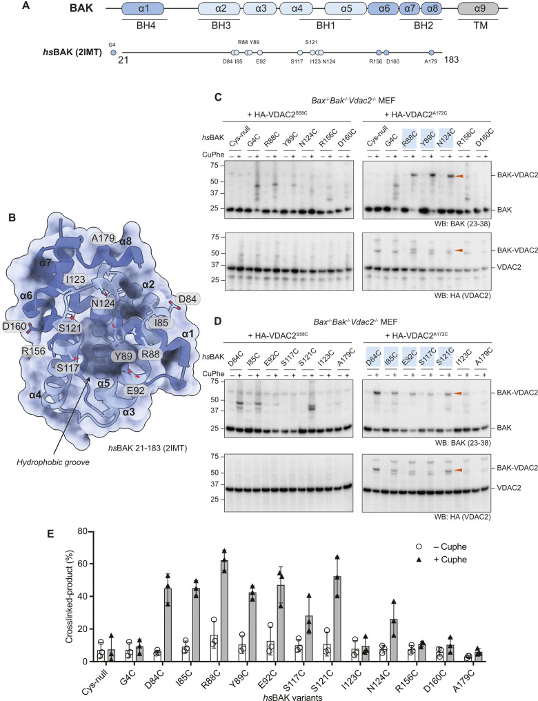
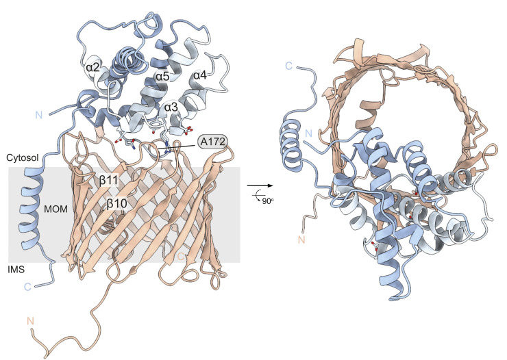
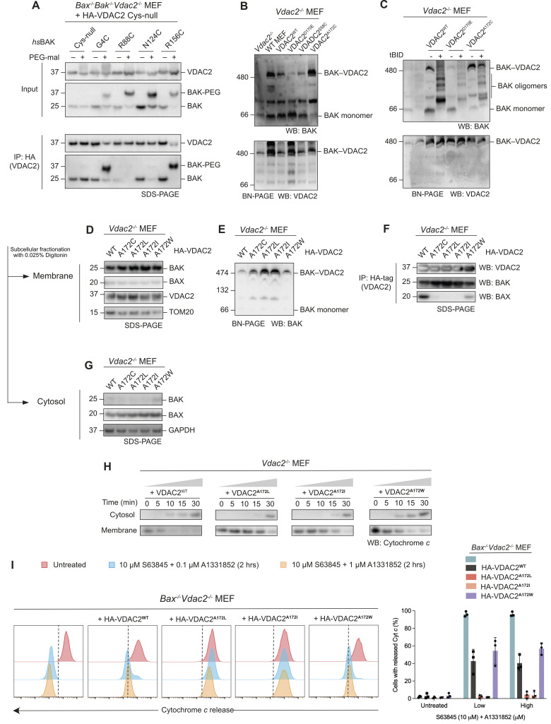
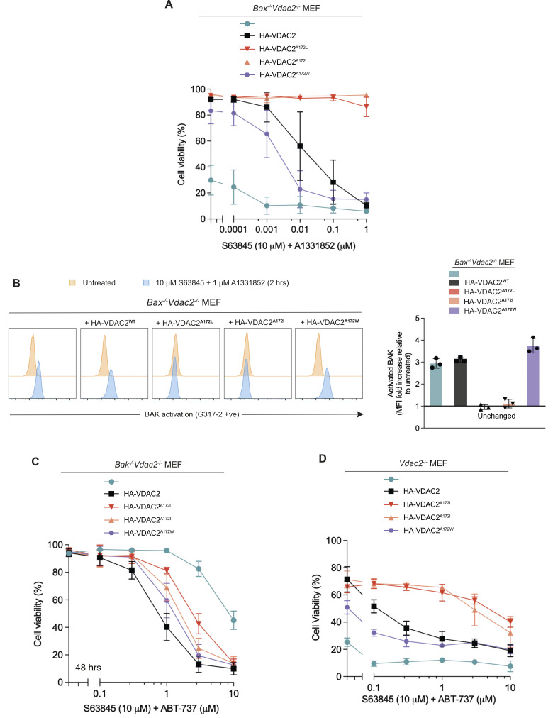
BAK and BAX execute intrinsic apoptosis by permeabilising the mitochondrial outer membrane. Their activity is regulated through interactions with pro-survival BCL-2 family proteins and with non-BCL-2 proteins including the mitochondrial channel protein VDAC2. VDAC2 is important for bringing both BAK and BAX to mitochondria where they execute their apoptotic function. Despite this important function in apoptosis, while interactions with pro-survival family members are well characterised and have culminated in the development of drugs that target these interfaces to induce cancer cell apoptosis, the interaction between BAK and VDAC2 remains largely undefined. Deep scanning mutagenesis coupled with cysteine linkage identified key residues in the interaction between BAK and VDAC2. Obstructive labelling of specific residues in the BH3 domain or hydrophobic groove of BAK disrupted this interaction. Conversely, mutating specific residues in a cytosol-exposed region of VDAC2 stabilised the interaction with BAK and inhibited BAK apoptotic activity. Thus, this VDAC2-BAK interaction site can potentially be targeted to either inhibit BAK-mediated apoptosis in scenarios where excessive apoptosis contributes to disease or to promote BAK-mediated apoptosis for cancer therapy.
BAK 和 BAX 通过使线粒体外膜通透来执行内在凋亡。它们的活性通过与生存促进 BCL-2 家族蛋白的相互作用以及与非 BCL-2 蛋白(包括线粒体通道蛋白 VDAC2)的相互作用来调节。VDAC2 对于将 BAK 和 BAX 带到线粒体中使其执行凋亡功能非常重要。尽管在凋亡中具有重要功能,但尽管与生存促进家族成员的相互作用已经得到很好的描述,并最终开发出靶向这些界面以诱导癌细胞凋亡的药物,但 BAK 和 VDAC2 之间的相互作用在很大程度上仍未得到定义。深扫描诱变结合半胱氨酸连接鉴定了 BAK 和 VDAC2 之间相互作用的关键残基。BAK 的 BH3 结构域或疏水性槽中特定残基的阻塞标记破坏了这种相互作用。相反,突变 VDAC2 细胞质暴露区域中的特定残基稳定了与 BAK 的相互作用并抑制了 BAK 的凋亡活性。因此,这个 VDAC2-BAK 相互作用位点可以潜在地靶向抑制在过度凋亡导致疾病的情况下 BAK 介导的凋亡,或者促进 BAK 介导的凋亡用于癌症治疗。