Osawa Iichiro, Mitsufuji Takashi, Nagawa Keita, Hara Yuki, Yamamoto Toshimasa, Araki Nobuo, Kozawa Eito
Department of Radiology, Saitama Medical University Hospital, 38 Morohongo, Moroyama-machi, Iruma-gun, Saitama 350-0495, Japan.
Department of Neurology, Saitama Medical University Hospital, 38 Morohongo, Moroyama-machi, Iruma-gun, Saitama 350-0495, Japan.
Eur J Radiol Open. 2024 Apr 25;12:100565. doi: 10.1016/j.ejro.2024.100565. eCollection 2024 Jun.
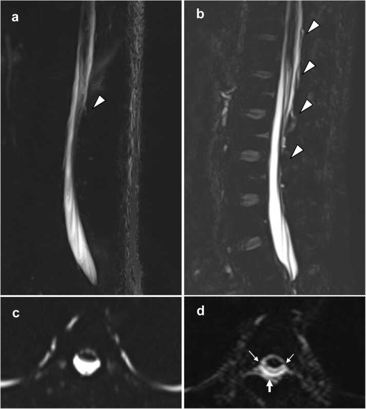
We compared cerebrospinal fluid (CSF) leak conspicuity and image quality as visualized using 3D versus 2D magnetic resonance (MR) myelography in patients with spinal CSF leaks.
Eighteen patients underwent spinal MR imaging at 3 Tesla. Three board-certified radiologists independently evaluated CSF leak conspicuity and image quality on a 4-point scale; the latter assessed by scoring fat suppression, venous visualization, and severity of CSF flow artifacts. Additionally, the evaluators ranked the overall performances of 2D versus 3D MR myelography upon completing side-by-side comparisons of CSF leak conspicuity. Inter-reader agreement was determined using the Gwet's AC1.
The quality of 3D MR myelography images was significantly better than that of 2D MR myelography with respect to CSF leak conspicuity (mean scores: 3.3 vs. 1.9, < 0.0001) and severity of CSF flow artifacts on the axial view (mean scores: 1.0 vs. 2.5, = 0.0001). Inter-reader agreement was moderate to almost perfect for 2D MR myelography (AC1 = 0.55-1.00), and almost perfect for 3D MR myelography (AC1 = 0.85-1.00). Moreover, 3D MR myelography was judged to be superior to 2D acquisition in 78 %, 83 %, and 83 % of the samples per readers 1, 2 and 3, respectively; the inter-reader agreement was almost perfect (AC1: reader 1 vs. 2; 0.98, reader 2 vs. 3; 0.96, reader 3 vs. 1; 0.98).
CSF leaks are more conspicuous when using 3D MR myelography than when using its 2D counterpart; therefore, the former is more reliable for identifying such leaks.
我们比较了在脊髓脑脊液漏患者中,使用三维(3D)与二维(2D)磁共振(MR)脊髓造影时脑脊液漏的显见度和图像质量。
18例患者在3特斯拉场强下接受脊髓MR成像。三位具有专业资格认证的放射科医生独立地按4分制评估脑脊液漏的显见度和图像质量;后者通过对脂肪抑制、静脉显影和脑脊液流动伪影的严重程度进行评分来评估。此外,评估者在对脑脊液漏显见度进行并排比较后,对2D与3D MR脊髓造影的总体表现进行排名。使用Gwet's AC1确定阅片者间的一致性。
在脑脊液漏显见度方面(平均得分:3.3对1.9,<0.0001)以及轴位视图上脑脊液流动伪影的严重程度方面(平均得分:1.0对2.5,=0.0001),3D MR脊髓造影图像的质量明显优于2D MR脊髓造影。2D MR脊髓造影的阅片者间一致性为中等至几乎完美(AC1 = 0.55 - 1.00),3D MR脊髓造影的阅片者间一致性几乎完美(AC1 = 0.85 - 1.00)。此外,对于阅片者1、2和3,分别有78%、83%和83%的样本判断3D MR脊髓造影优于2D采集;阅片者间一致性几乎完美(AC1:阅片者1对2;0.98,阅片者2对3;0.96,阅片者3对1;0.98)。
使用3D MR脊髓造影时脑脊液漏比使用其2D对应方法时更明显;因此,前者在识别此类漏液方面更可靠。