Okada Yukinori, Zama Tatsuhiko, Itonaga Tomohiro, Mikami Ryuji, Okubo Mitsuru, Sugahara Shinji, Nakai Motoki, Abe Koichiro, Yoshimura Mana, Saito Kazuhiro
Department of Radiology, Tokyo Medical University, Tokyo, Japan.
EJNMMI Rep. 2024 Feb 5;8(1):4. doi: 10.1186/s41824-024-00190-z.
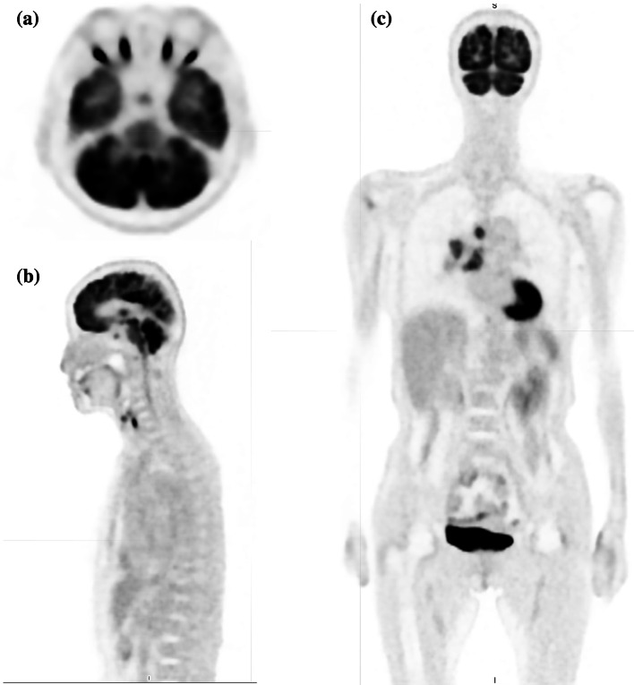
Research on the relationship between neuron-specific enolase (NSE) levels and normal organs, particularly the central nervous system, in small cell lung cancer is limited. Therefore, this study aimed to investigate the relationship between positron emission tomography-computed tomography (PET-CT) accumulation at hypothalamic/pituitary regions, tumor activity, and NSE level in limited-stage small cell lung cancer. We retrospectively analyzed patients who were diagnosed with limited-stage small cell lung cancer at Tokyo Medical University Hospital between July 1, 2019, and May 31, 2023, and were treated with chemoradiotherapy or radiotherapy. Leukocytes, erythrocytes, hemoglobin, platelets, total protein, albumin, NSE, and carcinoembryonic antigen were measured in blood samples obtained before treatment initiation. The maximum standardized uptake value (SUVmax), volume, and total lesion glycolysis (TLG) of each hypothalamic /pituitary region, primary tumor, and lymph node metastases were extracted from PET-CT images. The total tumor volume (primary tumor volume plus lymph node metastases volume) and total TLG (primary tumor TLG plus lymph node metastases TLG) were calculated.
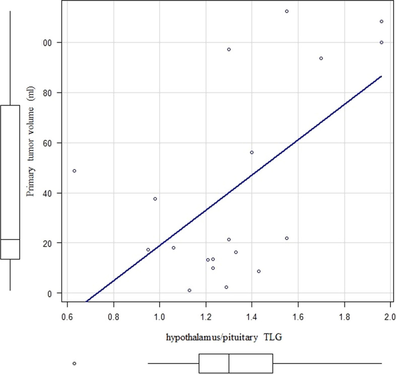
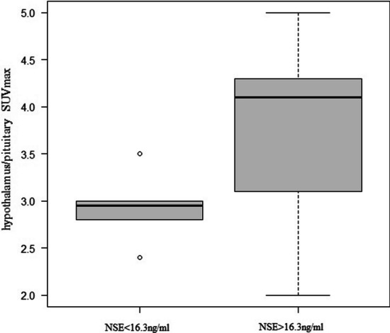
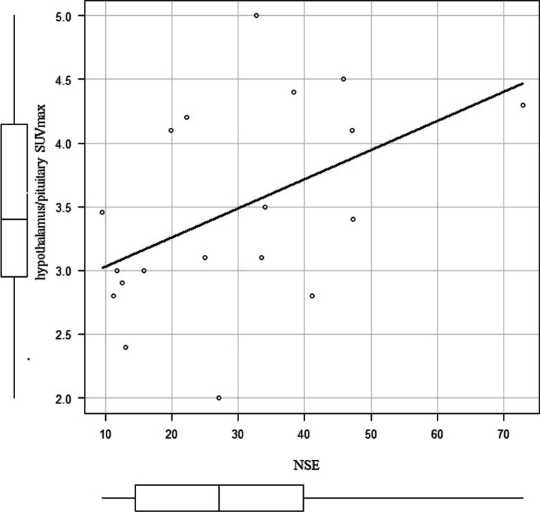
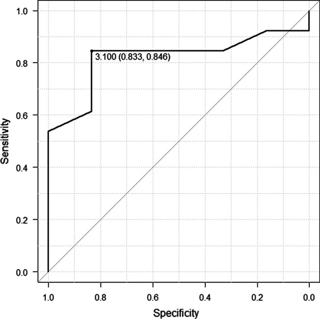
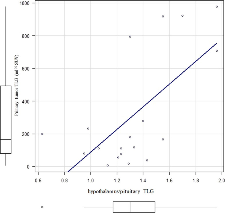
This study included 19 patients (mean age, 70.1 ± 8.8 years; 13 men and 6 women); the pathology in all patients was small cell lung cancer. Patients were classified into two groups according to the NSE reference value (16.3 ng/mL): six patients having NSE level below the reference value and 13 having NSE level above the reference value. The SUVmax in the hypothalamic/pituitary region was 2.95 in the NSE < 16.3 ng/mL group and 4.10 in the NSE > 16.3 ng/mL group, with a statistically significant difference (p = 0.03). The total tumor volume was 17.8 mL in the NSE < 16.3 ng/mL group and 98.9 mL in the NSE > 16.3 ng/mL group, with a statistically significant difference (p < 0.01). A correlation coefficient of r = 0.458 (p = 0.0486) was observed between SUVmax in the hypothalamus/pituitary and NSE level. A correlation coefficient of r = 0.647 (p < 0.01) was also observed between total tumor volume and NSE level. Finally, a correlation coefficient of r = 0.53 (p = 0.01) was observed between hypothalamic/pituitary TLG and primary tumor TLG.
The findings demonstrated a correlation between hypothalamic/pituitary activity and tumor activity, suggesting the prognostic significance of NSE.
关于小细胞肺癌中神经元特异性烯醇化酶(NSE)水平与正常器官,尤其是中枢神经系统之间关系的研究有限。因此,本研究旨在探讨局限期小细胞肺癌下丘脑/垂体区域的正电子发射断层扫描-计算机断层扫描(PET-CT)显像、肿瘤活性与NSE水平之间的关系。我们回顾性分析了2019年7月1日至2023年5月31日期间在东京医科大学医院被诊断为局限期小细胞肺癌并接受放化疗或放疗的患者。在开始治疗前采集的血样中检测白细胞、红细胞、血红蛋白、血小板、总蛋白、白蛋白、NSE和癌胚抗原。从PET-CT图像中提取每个下丘脑/垂体区域、原发肿瘤和淋巴结转移灶的最大标准化摄取值(SUVmax)、体积和总病灶糖酵解(TLG)。计算总肿瘤体积(原发肿瘤体积加淋巴结转移灶体积)和总TLG(原发肿瘤TLG加淋巴结转移灶TLG)。
本研究纳入19例患者(平均年龄70.1±8.8岁;男性13例,女性6例);所有患者的病理均为小细胞肺癌。根据NSE参考值(16.3 ng/mL)将患者分为两组:6例NSE水平低于参考值,13例NSE水平高于参考值。NSE<16.3 ng/mL组下丘脑/垂体区域的SUVmax为2.95,NSE>16.3 ng/mL组为4.10,差异有统计学意义(p = 0.03)。NSE<16.3 ng/mL组的总肿瘤体积为17.8 mL,NSE>16.3 ng/mL组为98.9 mL,差异有统计学意义(p<0.01)。下丘脑/垂体的SUVmax与NSE水平之间的相关系数r = 0.458(p = 0.0486)。总肿瘤体积与NSE水平之间也观察到相关系数r = 0.647(p<0.01)。最后,下丘脑/垂体TLG与原发肿瘤TLG之间的相关系数r = 0.53(p = 0.01)。
研究结果表明下丘脑/垂体活性与肿瘤活性之间存在相关性,提示NSE具有预后意义。