• 文献检索
  • 文档翻译
  • 深度研究
  • 学术资讯
  • Suppr Zotero 插件Zotero 插件
  • 邀请有礼
  • 套餐&价格
  • 历史记录
应用&插件
Suppr Zotero 插件Zotero 插件浏览器插件Mac 客户端Windows 客户端微信小程序
定价
高级版会员购买积分包购买API积分包
服务
文献检索文档翻译深度研究API 文档MCP 服务
关于我们
关于 Suppr公司介绍联系我们用户协议隐私条款
关注我们

Suppr 超能文献

核心技术专利:CN118964589B侵权必究
粤ICP备2023148730 号-1Suppr @ 2026

文献检索

告别复杂PubMed语法,用中文像聊天一样搜索,搜遍4000万医学文献。AI智能推荐,让科研检索更轻松。

立即免费搜索

文件翻译

保留排版,准确专业,支持PDF/Word/PPT等文件格式,支持 12+语言互译。

免费翻译文档

深度研究

AI帮你快速写综述,25分钟生成高质量综述,智能提取关键信息,辅助科研写作。

立即免费体验

肾上腺洗脱 CT 在无癌症病史患者中的应用:浪费时间吗?

Adrenal washout CT in patients with no history of cancer: a waste of time?

机构信息

Sir Charles Gairdner Hospital, Nedlands, WA, Australia.

Department of Research, Sir Charles Gairdner Hospital, Nedlands, WA, Australia.

出版信息

Abdom Radiol (NY). 2024 Sep;49(9):3117-3126. doi: 10.1007/s00261-024-04333-5. Epub 2024 May 21.

DOI:10.1007/s00261-024-04333-5
PMID:38772953
原文链接:https://pmc.ncbi.nlm.nih.gov/articles/PMC11335932/
Abstract

PURPOSE

To validate the diagnostic performance of adrenal washout CT in patients without known malignancy in a Western Australian population.

METHODS

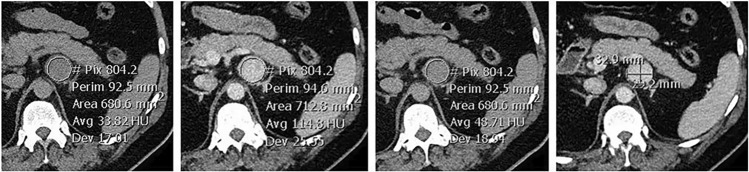
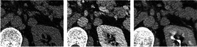
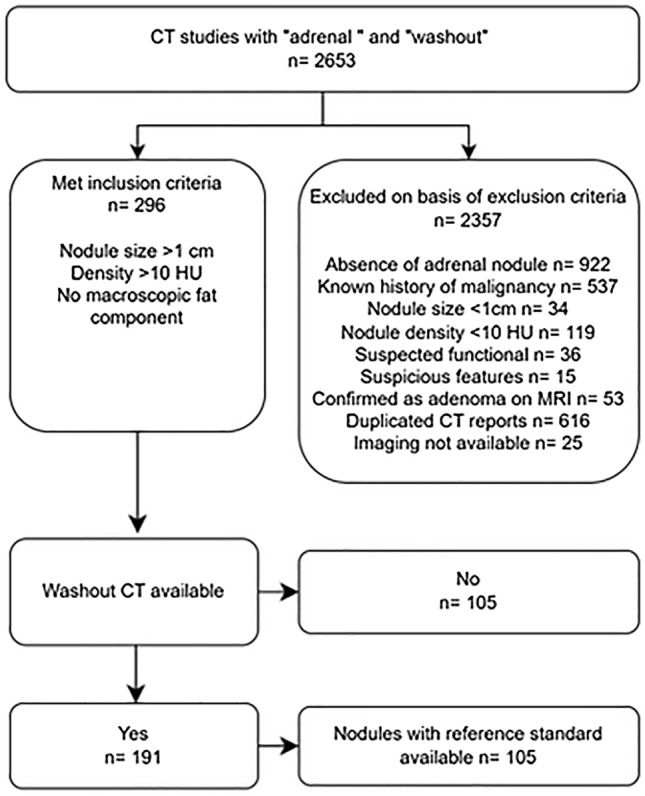
A radiology information system (RIS) search for CT reports containing "adrenal" and "washout" across six networked metropolitan public hospitals between January 2005 and November 2021. Homogenous nodules ≥ 1 cm, ≥ 10 HU without a suspected functional component in patients without a history of malignancy were included. Reported absolute and relative washout percentages were recorded and re-measured from unenhanced, 60-s portal venous and 15-min delayed phase imaging and compared to either histopathological or CT follow up for growth (≥ 12 months) reference standards.

RESULTS

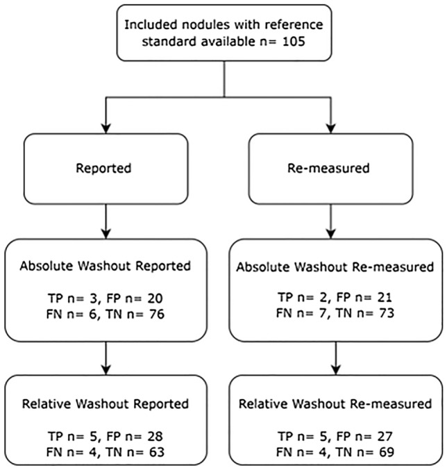
2653 studies were screened with 191 meeting inclusion criteria. 105 nodules underwent washout CT and then had either histopathological (12 patients) or CT follow up (93 patients) reference standards available. Reported absolute washout (aWO) estimated sensitivity and specificity for malignant/indeterminate nodules was low at 33% (95% CI 25-43%) and 77% (95% CI 68-84%) respectively. Reported relative washout (rWO) sensitivity and specificity were 56% (95% CI 46-65%) and 69% (95% CI 60-77%) respectively. Negative predictive values for both aWO and rWO were reassuring at 92% (95% CI 86-96%) and 94% (95%CI 88-97%).

CONCLUSION

Our study validates a recent report suggesting that adrenal washout has poor sensitivity for and consequent limited utility to exclude malignancy in patients with no cancer history. However, patients with incidental adrenal nodules < 4 cm in size with benign washout can be reassured by the high negative predictive value and worked up to exclude functional adenoma and re-imaged in a year to confirm no growth.

摘要

目的

在西澳大利亚人群中验证无已知恶性肿瘤的患者中肾上腺洗脱 CT 的诊断性能。

方法

对 2005 年 1 月至 2021 年 11 月期间在六家联网的大都市公立医院的 CT 报告进行放射学信息系统 (RIS) 搜索,以查找包含“肾上腺”和“洗脱”的 CT 报告。纳入无恶性肿瘤病史且均质结节≥1cm,≥10HU 且无可疑功能成分的患者。记录报告的绝对洗脱率和相对洗脱率,并从未增强、60 秒门静脉期和 15 分钟延迟期成像中重新测量,并与组织病理学或 CT 随访的生长(≥12 个月)参考标准进行比较。

结果

共筛选了 2653 项研究,其中 191 项符合纳入标准。105 个结节进行了洗脱 CT 检查,然后有组织病理学(12 例)或 CT 随访(93 例)参考标准。报告的恶性/不确定结节的绝对洗脱(aWO)估计敏感性和特异性分别为 33%(95%CI 25-43%)和 77%(95%CI 68-84%)。报告的相对洗脱(rWO)的敏感性和特异性分别为 56%(95%CI 46-65%)和 69%(95%CI 60-77%)。aWO 和 rWO 的阴性预测值分别为 92%(95%CI 86-96%)和 94%(95%CI 88-97%),令人放心。

结论

我们的研究验证了最近的一项报告,即对于无癌症病史的患者,肾上腺洗脱对恶性肿瘤的敏感性差,因此对排除恶性肿瘤的作用有限。然而,对于大小<4cm 的偶然发现的肾上腺结节且洗脱良性的患者,可以通过高阴性预测值来安心,并排除功能性腺瘤并在一年内重新成像以确认无生长。

https://cdn.ncbi.nlm.nih.gov/pmc/blobs/ad40/11335932/1facef14f51c/261_2024_4333_Fig5_HTML.jpg
https://cdn.ncbi.nlm.nih.gov/pmc/blobs/ad40/11335932/5abf001c7920/261_2024_4333_Fig1_HTML.jpg
https://cdn.ncbi.nlm.nih.gov/pmc/blobs/ad40/11335932/b6bb11e14ea4/261_2024_4333_Fig2_HTML.jpg
https://cdn.ncbi.nlm.nih.gov/pmc/blobs/ad40/11335932/0c1ea7e29a3b/261_2024_4333_Fig3_HTML.jpg
https://cdn.ncbi.nlm.nih.gov/pmc/blobs/ad40/11335932/09c297c9ebc9/261_2024_4333_Fig4_HTML.jpg
https://cdn.ncbi.nlm.nih.gov/pmc/blobs/ad40/11335932/1facef14f51c/261_2024_4333_Fig5_HTML.jpg

相似文献

1
Adrenal washout CT in patients with no history of cancer: a waste of time?肾上腺洗脱 CT 在无癌症病史患者中的应用:浪费时间吗?
Abdom Radiol (NY). 2024 Sep;49(9):3117-3126. doi: 10.1007/s00261-024-04333-5. Epub 2024 May 21.
2
Incidental Adrenal Nodules in Patients Without Known Malignancy: Prevalence of Malignancy and Utility of Washout CT for Characterization-A Multiinstitutional Study.无已知恶性肿瘤的患者偶然发现的肾上腺结节:恶性肿瘤的患病率和洗脱 CT 对特征描述的效用-一项多机构研究。
AJR Am J Roentgenol. 2022 Nov;219(5):804-812. doi: 10.2214/AJR.22.27901. Epub 2022 Jun 22.
3
Performance of CT With Adrenal-Washout Protocol in Heterogeneous Adrenal Nodules: A Multiinstitutional Study.CT 肾上腺洗脱方案在异质性肾上腺结节中的应用性能:一项多机构研究。
AJR Am J Roentgenol. 2024 May;222(5):e2330769. doi: 10.2214/AJR.23.30769. Epub 2024 May 29.
4
Adrenal Adenoma and Pheochromocytoma: Comparison of Multidetector CT Venous Enhancement Levels and Washout Characteristics.肾上腺腺瘤与嗜铬细胞瘤:多排CT静脉强化水平及廓清特征比较
J Comput Assist Tomogr. 2016 Mar-Apr;40(2):194-200. doi: 10.1097/RCT.0000000000000343.
5
Histogram analysis for characterization of indeterminate adrenal nodules on noncontrast CT.非增强CT上不确定肾上腺结节特征的直方图分析
Abdom Imaging. 2015 Aug;40(6):1666-74. doi: 10.1007/s00261-014-0307-6.
6
CT sensitivity for adrenal adenoma according to lesion size.根据病变大小的肾上腺腺瘤的CT敏感性。
Abdom Imaging. 2015 Oct;40(8):3152-60. doi: 10.1007/s00261-015-0521-x.
7
Differentiation of Adrenal Hyperplasia From Adenoma by Use of CT Densitometry and Percentage Washout.利用CT密度测定法和洗脱百分比鉴别肾上腺增生与腺瘤
AJR Am J Roentgenol. 2016 Jan;206(1):106-12. doi: 10.2214/AJR.15.14558.
8
Adenoma characterization: adrenal protocol with dual-energy CT.腺瘤特征描述:双能 CT 肾上腺议定书。
Radiology. 2013 Apr;267(1):155-63. doi: 10.1148/radiol.12112735. Epub 2013 Jan 17.
9
The value of 15-minute delayed contrast-enhanced CT to differentiate hyperattenuating adrenal masses compared with chemical shift MR imaging.与化学位移磁共振成像相比,15 分钟延迟对比增强 CT 对高信号肾上腺肿块的鉴别价值。
Eur Radiol. 2014 Jun;24(6):1410-20. doi: 10.1007/s00330-013-3084-7. Epub 2014 Mar 20.
10
Pheochromocytomas Versus Adenoma: Role of Venous Phase CT Enhancement.嗜铬细胞瘤与腺瘤:静脉期 CT 增强的作用。
AJR Am J Roentgenol. 2018 May;210(5):1073-1078. doi: 10.2214/AJR.17.18472. Epub 2018 Mar 23.

引用本文的文献

1
Washed up: the end of an era for adrenal incidentaloma CT.大势已去:肾上腺偶发瘤CT检查时代的终结
Insights Imaging. 2025 Jun 27;16(1):136. doi: 10.1186/s13244-025-02015-4.
2
Feasibility of extracellular volume fraction derived from single-phase spectral CT for differentiating between adrenal metastases and adenomas.基于单相光谱CT得出的细胞外体积分数在鉴别肾上腺转移瘤和腺瘤中的可行性。
Abdom Radiol (NY). 2025 May;50(5):2160-2168. doi: 10.1007/s00261-024-04681-2. Epub 2024 Nov 8.

本文引用的文献

1
Approach to the Patient With Adrenal Hemorrhage.肾上腺出血患者的处理方法。
J Clin Endocrinol Metab. 2023 Mar 10;108(4):995-1006. doi: 10.1210/clinem/dgac672.
2
Incidental Adrenal Nodules in Patients Without Known Malignancy: Prevalence of Malignancy and Utility of Washout CT for Characterization-A Multiinstitutional Study.无已知恶性肿瘤的患者偶然发现的肾上腺结节:恶性肿瘤的患病率和洗脱 CT 对特征描述的效用-一项多机构研究。
AJR Am J Roentgenol. 2022 Nov;219(5):804-812. doi: 10.2214/AJR.22.27901. Epub 2022 Jun 22.
3
A Critical Analysis of Computed Tomography Washout in Lipid-Poor Adrenal Incidentalomas.
对乏脂性肾上腺偶发瘤的计算机断层扫描洗脱的批判性分析
Ann Surg Oncol. 2021 May;28(5):2756-2762. doi: 10.1245/s10434-020-09329-1. Epub 2020 Nov 18.
4
Adrenal Incidentaloma.肾上腺意外瘤。
Endocr Rev. 2020 Dec 1;41(6):775-820. doi: 10.1210/endrev/bnaa008.
5
Differences in Growth Rate on CT of Adrenal Adenomas and Malignant Adrenal Nodules.肾上腺腺瘤和恶性肾上腺结节 CT 生长速度的差异。
AJR Am J Roentgenol. 2019 Sep;213(3):632-636. doi: 10.2214/AJR.19.21342. Epub 2019 Apr 30.
6
Imaging features of adrenal masses.肾上腺肿块的影像学特征。
Insights Imaging. 2019 Jan 25;10(1):1. doi: 10.1186/s13244-019-0688-8.
7
Management of Incidental Adrenal Masses: A White Paper of the ACR Incidental Findings Committee.偶发肾上腺肿块的管理:美国放射学会偶发发现委员会白皮书
J Am Coll Radiol. 2017 Aug;14(8):1038-1044. doi: 10.1016/j.jacr.2017.05.001. Epub 2017 Jun 23.
8
Management of adrenal incidentalomas: European Society of Endocrinology Clinical Practice Guideline in collaboration with the European Network for the Study of Adrenal Tumors.肾上腺偶发瘤的管理:欧洲内分泌学会临床实践指南与欧洲肾上腺肿瘤研究网络合作制定
Eur J Endocrinol. 2016 Aug;175(2):G1-G34. doi: 10.1530/EJE-16-0467.
9
Adrenal Incidentalomas: Clinical Controversies and Modified Recommendations.肾上腺偶发瘤:临床争议与修订建议
AJR Am J Roentgenol. 2016 Jun;206(6):1170-8. doi: 10.2214/AJR.15.15475. Epub 2016 Apr 12.
10
Comparison of Quantitative MRI and CT Washout Analysis for Differentiation of Adrenal Pheochromocytoma From Adrenal Adenoma.定量MRI与CT洗脱分析在鉴别肾上腺嗜铬细胞瘤与肾上腺腺瘤中的比较
AJR Am J Roentgenol. 2016 Jun;206(6):1141-8. doi: 10.2214/AJR.15.15318. Epub 2016 Mar 24.