Neuroradiology, Department of Neuroscience, University of Padua, Padua, Italy.
Dipartimento di Medicina e Chirurgia, Scuola Medica Salernitana, Università di Salerno, Fisciano, Italy.
Orphanet J Rare Dis. 2024 May 21;19(1):212. doi: 10.1186/s13023-024-03194-x.
Brain injury in hereditary hemoglobinopathies is commonly attributed to anemia-related relative hypoperfusion in terms of impaired oxygen blood supply. Supratentorial and infratentorial vascular watershed regions seem to be especially vulnerable, but data are very scarce.
We investigated a large beta-thalassemia sample with arterial spin labeling in order to characterize regional perfusion changes and their correlation with phenotype and anemia severity.
We performed a multicenter single-scanner cross-sectional 3T-MRI study analyzing non-invasively the brain perfusion in 54 transfusion-dependent thalassemia (TDT), 23 non-transfusion-dependent thalassemia (NTDT) patients and 56 Healthy Controls (HC). Age, hemoglobin levels, and cognitive functioning were recorded.
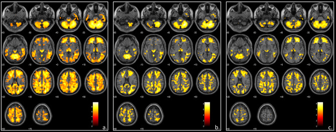
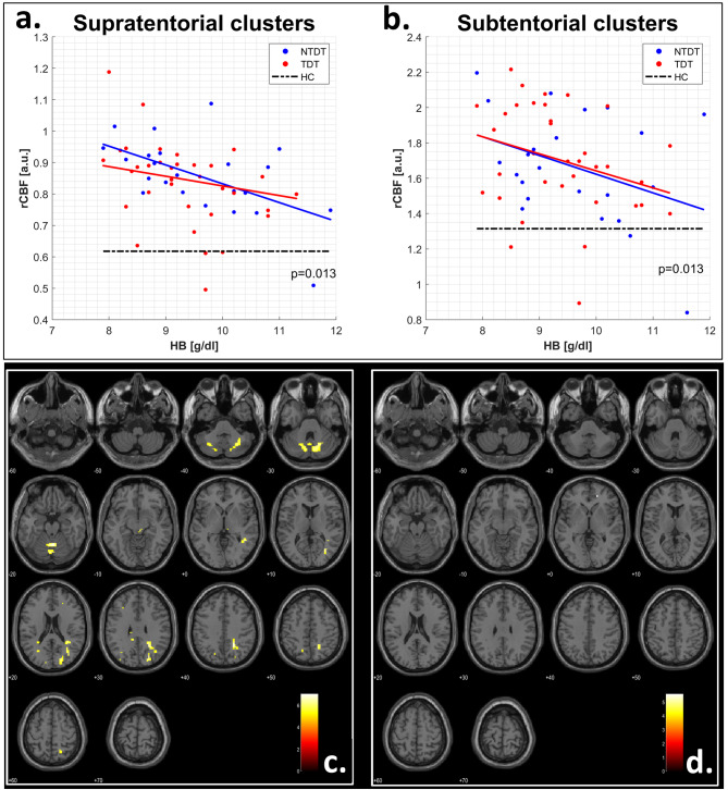
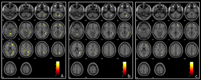
Both TDT and NTDT patients showed globally increased brain perfusion values compared to healthy controls, while no difference was found between patient subgroups. Using age and sex as covariates and scaling the perfusion maps for the global cerebral blood flow, beta-thalassemia patients showed relative hyperperfusion in supratentorial/infratentorial watershed regions. Perfusion changes correlated with hemoglobin levels (p = 0.013) and were not observed in the less severely anemic patients (hemoglobin level > 9.5 g/dL). In the hyperperfused regions, white matter density was significantly decreased (p = 0.0003) in both patient subgroups vs. HC. In NTDT, white matter density changes correlated inversely with full-scale Intelligence Quotient (p = 0.007) while in TDT no correlation was found.
Relative hyperperfusion of watershed territories represents a hemodynamic hallmark of beta-thalassemia anemia challenging previous hypotheses of brain injury in hereditary anemias. A careful management of anemia severity might be crucial for preventing structural white matter changes and subsequent long-term cognitive impairment.
遗传性血红蛋白病中的脑损伤通常归因于贫血相关的相对低灌注,表现为氧合血液供应受损。幕上和幕下血管分水岭区域似乎特别容易受到影响,但数据非常有限。
我们通过动脉自旋标记研究了一个大型的β-地中海贫血样本,以描述区域性灌注变化及其与表型和贫血严重程度的相关性。
我们进行了一项多中心单扫描仪 3T-MRI 横断面研究,分析了 54 例输血依赖性地中海贫血(TDT)、23 例非输血依赖性地中海贫血(NTDT)患者和 56 例健康对照(HC)的脑灌注情况。记录年龄、血红蛋白水平和认知功能。
TDT 和 NTDT 患者的脑灌注值均高于健康对照组,而患者亚组之间无差异。使用年龄和性别作为协变量,并对灌注图进行全局脑血流缩放,β-地中海贫血患者在幕上/幕下分水岭区域显示相对高灌注。灌注变化与血红蛋白水平相关(p=0.013),在贫血程度较轻的患者中未观察到(血红蛋白水平>9.5 g/dL)。在高灌注区域,两组患者的白质密度均显著降低(p=0.0003 与 HC 相比)。在 NTDT 中,白质密度变化与全量表智商呈负相关(p=0.007),而在 TDT 中未发现相关性。
分水岭区域的相对高灌注代表了β-地中海贫血贫血的血液动力学特征,挑战了先前关于遗传性贫血性脑损伤的假说。对贫血严重程度的仔细管理对于预防结构白质变化和随后的长期认知障碍可能至关重要。