Department of Veterinary Clinical Sciences, College of Veterinary Medicine, Purdue University, West Lafayette, Indiana, USA.
Veterinary Administration, College of Veterinary Medicine, Purdue University, West Lafayette, Indiana, USA.
J Vet Intern Med. 2024 Jul-Aug;38(4):2204-2213. doi: 10.1111/jvim.17109. Epub 2024 May 28.
The prognosis of individual dogs with meningoencephalomyelitis of unknown etiology (MUE) remains difficult to predict. MUE cases with no lesions detected by magnetic resonance imaging (MRI) occur, but it is unknown whether this finding is associated with prognosis.
MUE cases without detectable lesions on MRI have a better outcome than cases with detectable lesions.
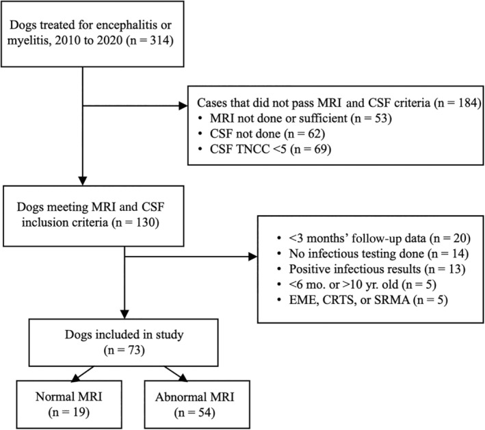
Study included 73 client-owned dogs with MUE presenting to Purdue University Veterinary Hospital from 2010 to 2020.
Retrospective study. Dogs with a clinical diagnosis of MUE were identified by medical record search. MRI reports were reviewed for presence or absence of lesions consistent with MUE. Clinical findings at presentation, treatment, disease-specific survival, and outcomes including rates of remission and relapse were compared between cases with normal MRI or abnormal MRI.
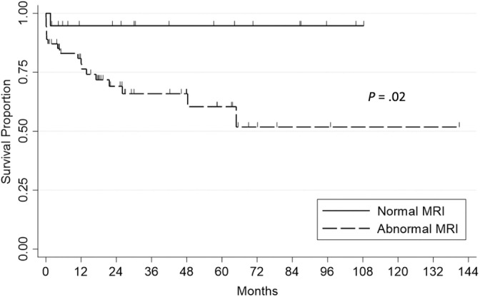
Overall, 54 dogs (74%) were classified as abnormal MRI, and 19 dogs (26%) were classified as normal MRI cases. Death caused by MUE occurred in 1/19 (5%) normal MRI dogs and 18/54 (33%) abnormal MRI dogs (P = .016). Median survival was >107 months in both groups, but survival was significantly longer in the normal MRI group (P = .019). On multivariate analysis, abnormal MRI was significantly related to death (hazard ratio, 7.71; 95% confidence interval 1.03-58.00, P = .0470), whereas significant relationships with death were not identified for either the use of secondary immunosuppressive medications or cerebrospinal fluid nucleated cell count.
MUE dogs with no detectable lesions on MRI have reduced disease-related death compared with dogs with abnormal MRI. The presence or absence of MRI lesions in MUE dogs is prognostically relevant.
个别病因不明的脑膜脑炎(MUE)犬的预后仍然难以预测。尽管磁共振成像(MRI)未发现病变,但仍会出现 MUE 病例,但目前尚不清楚这一发现是否与预后相关。
MRI 未检测到病变的 MUE 病例比可检测到病变的病例预后更好。
本研究纳入了 2010 年至 2020 年期间在普渡大学兽医院就诊的 73 例患有 MUE 的患犬。
回顾性研究。通过病历检索确定 MUE 的临床诊断。对 MRI 报告进行审查,以确定是否存在与 MUE 一致的病变。比较 MRI 正常和 MRI 异常的病例在就诊时的临床发现、治疗、疾病特异性生存以及包括缓解和复发率在内的结局。
总体而言,54 例(74%)犬被归类为 MRI 异常,19 例(26%)犬被归类为 MRI 正常。MRI 正常的 19 例犬中有 1 例(5%)死于 MUE,MRI 异常的 54 例犬中有 18 例(33%)死于 MUE(P = .016)。两组的中位生存时间均超过 107 个月,但 MRI 正常组的生存时间明显更长(P = .019)。多变量分析显示,MRI 异常与死亡显著相关(风险比,7.71;95%置信区间,1.03-58.00,P =.0470),而 secondary 免疫抑制药物或脑脊液有核细胞计数与死亡无显著关系。
MRI 未检测到病变的 MUE 犬与 MRI 异常犬相比,疾病相关死亡率降低。MUE 犬 MRI 病变的存在与否与预后相关。