• 文献检索
  • 文档翻译
  • 深度研究
  • 学术资讯
  • Suppr Zotero 插件Zotero 插件
  • 邀请有礼
  • 套餐&价格
  • 历史记录
应用&插件
Suppr Zotero 插件Zotero 插件浏览器插件Mac 客户端Windows 客户端微信小程序
定价
高级版会员购买积分包购买API积分包
服务
文献检索文档翻译深度研究API 文档MCP 服务
关于我们
关于 Suppr公司介绍联系我们用户协议隐私条款
关注我们

Suppr 超能文献

核心技术专利:CN118964589B侵权必究
粤ICP备2023148730 号-1Suppr @ 2026

文献检索

告别复杂PubMed语法,用中文像聊天一样搜索,搜遍4000万医学文献。AI智能推荐,让科研检索更轻松。

立即免费搜索

文件翻译

保留排版,准确专业,支持PDF/Word/PPT等文件格式,支持 12+语言互译。

免费翻译文档

深度研究

AI帮你快速写综述,25分钟生成高质量综述,智能提取关键信息,辅助科研写作。

立即免费体验

俯卧位胸腔镜下食管球瘤摘除术:1例报告及文献复习

Thoracoscopic enucleation of an esophageal glomus tumor in the prone position: a case report and literature review.

作者信息

Matsumoto Shigeki, Okumura Tomoyuki, Miwa Takeshi, Numata Yoshihisa, Hamashima Takeru, Ito Miki, Nagaoka Yasuhiro, Takeshita Chitaru, Sakai Ayano, Kimura Nana, Fukasawa Mina, Mori Kosuke, Takeda Naoya, Yagi Kenta, Muranushi Ryo, Manabe Takahiro, Shirai Yoshihiro, Watanabe Toru, Hirano Katsuhisa, Hashimoto Isaya, Shibuya Kazuto, Yoshioka Isaku, Fujii Tsutomu

机构信息

Department of Surgery and Science, Faculty of Medicine, Academic Assembly, University of Toyama, 2630 Sugitani, Toyama, 930-0194, Japan.

Office of Human Research Ethics, Faculty of Education and Research Promotion, Academic Assembly, University of Toyama, 2630 Sugitani, Toyama, 930-0194, Japan.

出版信息

Surg Case Rep. 2024 May 28;10(1):131. doi: 10.1186/s40792-024-01934-6.

DOI:10.1186/s40792-024-01934-6
PMID:38805072
原文链接:https://pmc.ncbi.nlm.nih.gov/articles/PMC11133250/
Abstract

BACKGROUND

Glomus tumors (GT) generally occur in the skin. However, esophageal GT, an extremely rare condition, has no established standardized treatment guidelines. Herein, we report the case of an esophageal GT successfully removed by thoracoscopic enucleation in the prone position using intra-esophageal balloon compression.

CASE PRESENTATION

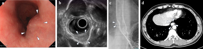
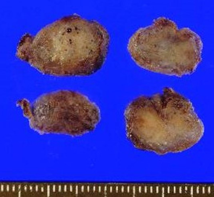
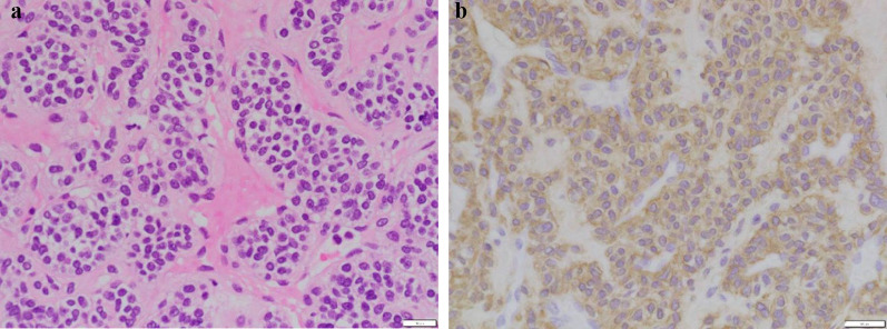
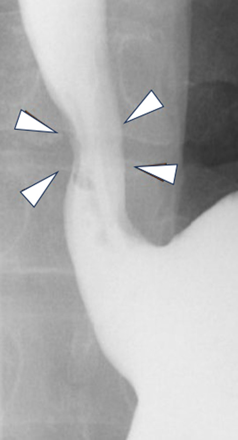
A 45-year-old man underwent an annual endoscopic examination and was found to have a submucosal tumor in the lower esophagus. Endoscopic ultrasound (EUS) revealed a hyperechoic mass originating from the muscular layer. Contrast-enhanced computed tomography identified a 2 cm mass lesion with high contrast enhancement in the right side of the lower esophagus. Pathologic findings of EUS-guided fine needle aspiration biopsy (EUS-FNA) revealed round to spindle shaped atypical cells without mitotic activity. Immunohistochemically, the tumor was positive for alpha-smooth muscle actin, but negative for CD34, desmin, keratin 18, S-100 protein, melan A, c-kit, and STAT6. He was diagnosed with an esophageal GT and a thoracoscopic approach to tumor resection was planned. Under general anesthesia, a Sengstaken-Blakemore (SB) tube was inserted into the esophagus. The patient was placed in the prone position and a right thoracoscopic approach was achieved. The esophagus around the tumor was mobilized and the SB tube balloon inflated to compress the tumor toward the thoracic cavity. The muscle layer was divided and the tumor was successfully enucleated without mucosal penetration. Oral intake was initiated on postoperative day (POD) 3 and the patient discharged on POD 9. No surgical complications or tumor metastasis were observed during the 1-year postoperative follow-up.

CONCLUSIONS

As malignancy criteria for esophageal GT are not yet established, the least invasive procedure for complete resection should be selected on a case-by-case basis. Thoracoscopic enucleation in the prone position using intra-esophageal balloon compression is useful to treat esophageal GT on the right side of the esophagus.

摘要

背景

血管球瘤(GT)通常发生于皮肤。然而,食管GT极为罕见,尚无既定的标准化治疗指南。在此,我们报告一例通过胸腔镜在俯卧位使用食管内球囊压迫成功摘除的食管GT病例。

病例介绍

一名45岁男性接受年度内镜检查时,发现食管下段有一黏膜下肿瘤。内镜超声(EUS)显示一个起源于肌层的高回声肿块。增强计算机断层扫描显示食管下段右侧有一个2厘米的肿块病变,具有高对比度增强。EUS引导下细针穿刺活检(EUS-FNA)的病理结果显示为圆形至梭形的非典型细胞,无有丝分裂活性。免疫组织化学检查显示,肿瘤α-平滑肌肌动蛋白呈阳性,但CD34、结蛋白、细胞角蛋白18、S-100蛋白、黑素A、c-kit和信号转导和转录激活因子6呈阴性。他被诊断为食管GT,并计划采用胸腔镜方法进行肿瘤切除。在全身麻醉下,将一根森斯塔肯-布莱克莫尔(SB)管插入食管。患者置于俯卧位,采用右侧胸腔镜入路。游离肿瘤周围的食管,充盈SB管球囊以将肿瘤压向胸腔。切开肌层,成功摘除肿瘤,未穿透黏膜。术后第3天开始经口进食,患者于术后第9天出院。术后1年随访期间未观察到手术并发症或肿瘤转移。

结论

由于食管GT的恶性标准尚未确立,应根据具体情况选择创伤最小的完整切除手术。胸腔镜在俯卧位使用食管内球囊压迫对治疗食管右侧的GT是有用的。

https://cdn.ncbi.nlm.nih.gov/pmc/blobs/1344/11133250/153b46b0667a/40792_2024_1934_Fig4_HTML.jpg
https://cdn.ncbi.nlm.nih.gov/pmc/blobs/1344/11133250/bb45dfa9b57d/40792_2024_1934_Fig1_HTML.jpg
https://cdn.ncbi.nlm.nih.gov/pmc/blobs/1344/11133250/07a04a9587e3/40792_2024_1934_Fig2_HTML.jpg
https://cdn.ncbi.nlm.nih.gov/pmc/blobs/1344/11133250/c69aacd547c0/40792_2024_1934_Fig3_HTML.jpg
https://cdn.ncbi.nlm.nih.gov/pmc/blobs/1344/11133250/153b46b0667a/40792_2024_1934_Fig4_HTML.jpg

相似文献

1
Thoracoscopic enucleation of an esophageal glomus tumor in the prone position: a case report and literature review.俯卧位胸腔镜下食管球瘤摘除术:1例报告及文献复习
Surg Case Rep. 2024 May 28;10(1):131. doi: 10.1186/s40792-024-01934-6.
2
Thoracoscopic enucleation in the left decubitus position for leiomyoma of the upper thoracic esophagus: Utility of preoperative diagnosis applying endoscopic ultrasound-guided fine needle aspiration.左侧卧位胸腔镜下摘除术治疗胸段上段食管平滑肌瘤:应用超声内镜引导下细针穿刺进行术前诊断的效用
Int J Surg Case Rep. 2017;34:49-55. doi: 10.1016/j.ijscr.2017.03.007. Epub 2017 Mar 8.
3
Resection of an esophageal schwannoma with thoracoscopic surgery: a case report.胸腔镜手术切除食管神经鞘瘤:一例报告
Surg Case Rep. 2016 Dec;2(1):127. doi: 10.1186/s40792-016-0256-0. Epub 2016 Nov 7.
4
Thoracoscopic enucleation of esophageal stromal tumor.胸腔镜下食管间质瘤摘除术
Dis Esophagus. 2004;17(1):104-8. doi: 10.1111/j.1442-2050.2004.00364.x.
5
Giant circumferential esophageal leiomyoma successfully treated by thoracoscopic enucleation with the patient in a prone position: A case report.胸腔镜下俯卧位切除巨大环状食管平滑肌瘤 1 例报告
Asian J Endosc Surg. 2021 Jul;14(3):602-606. doi: 10.1111/ases.12910. Epub 2020 Dec 14.
6
Successful enucleation of a fluorine-18-fluorodeoxyglucose positron emission tomography positive esophageal leiomyoma in the prone position using sponge spacer and intra-esophageal balloon compression.使用海绵间隔物和食管内球囊压迫在俯卧位成功摘除氟-18-氟脱氧葡萄糖正电子发射断层扫描阳性的食管平滑肌瘤。
Gen Thorac Cardiovasc Surg. 2012 Aug;60(8):542-5. doi: 10.1007/s11748-012-0027-1. Epub 2012 May 8.
7
Asymptomatic esophageal glomus tumor: case report.无症状性食管血管球瘤:病例报告
J Gastrointest Oncol. 2019 Oct;10(5):1015-1020. doi: 10.21037/jgo.2019.05.08.
8
Thoracoscopic surgery combined with endoscopic creation of a submucosal tunnel for a large complicated esophageal leiomyoma.胸腔镜手术联合内镜下黏膜下隧道技术治疗巨大复杂性食管平滑肌瘤
Surg Case Rep. 2020 May 6;6(1):92. doi: 10.1186/s40792-020-00854-5.
9
[A Case of Thoracoscopic Tumor Enucleation of Esophageal Leiomyoma].[胸腔镜下食管平滑肌瘤摘除术 1 例]
Gan To Kagaku Ryoho. 2023 Mar;50(3):387-389.
10
Robotic-assisted enucleation of a large lower esophageal leiomyoma and review of literature.机器人辅助下的巨大食管下段平滑肌瘤切除术及文献复习。
Int J Med Robot. 2013 Sep;9(3):253-7. doi: 10.1002/rcs.1484. Epub 2013 Feb 12.

本文引用的文献

1
Gastric Glomus Tumors: The Roles of Endoscopic Ultrasound and Shared Decision-Making.胃血管球瘤:内镜超声及共同决策的作用
Case Rep Gastroenterol. 2023 Dec 8;17(1):356-361. doi: 10.1159/000534643. eCollection 2023 Jan-Dec.
2
Glomus Tumor of the Stomach: A Systematic Review and Illustrative Case Report.胃血管球瘤:系统评价及病例报告。
Dig Dis. 2023;41(1):17-33. doi: 10.1159/000525513. Epub 2022 Jun 26.
3
Rare malignant glomus tumor of the esophagus with pulmonary metastasis: a case report.罕见的食管恶性血管球瘤伴肺转移:一例报告
AME Case Rep. 2022 Apr 25;6:20. doi: 10.21037/acr-21-72. eCollection 2022.
4
Thoracoscopic enucleation of oesophageal submucosal tumours in prone position gives excellent long-term outcome: A single-centre experience.俯卧位胸腔镜下食管黏膜下肿瘤摘除术的长期效果极佳:单中心经验
J Minim Access Surg. 2022 Jul-Sep;18(3):401-407. doi: 10.4103/jmas.jmas_169_21.
5
Robot-assisted thoracoscopic enucleation for a large esophageal leiomyoma: a case report.机器人辅助胸腔镜下巨大食管平滑肌瘤摘除术:一例报告
Surg Case Rep. 2021 May 26;7(1):129. doi: 10.1186/s40792-021-01212-9.
6
Clinical Implications of 18F-FDG PET/CT in Malignant Glomus Tumors of the Esophagus.18F-FDG PET/CT 对食管恶性血管球瘤的临床意义。
Clin Nucl Med. 2020 Jun;45(6):e301-e302. doi: 10.1097/RLU.0000000000003029.
7
Asymptomatic esophageal glomus tumor: case report.无症状性食管血管球瘤:病例报告
J Gastrointest Oncol. 2019 Oct;10(5):1015-1020. doi: 10.21037/jgo.2019.05.08.
8
Glomus Tumor: Twenty-Year Experience and Literature Review.血管球瘤:二十年经验及文献综述
Ann Plast Surg. 2016 Mar;76 Suppl 1:S35-40. doi: 10.1097/SAP.0000000000000684.
9
Glomus Tumor of the Esophagus: A Case Report and Review of the Literature.食管球瘤:一例报告并文献复习
Conn Med. 2015 Feb;79(2):93-5.
10
Malignant glomus tumor with oncocytic features: an unusual presentation of dysphagia.具有嗜酸性细胞特征的恶性血管球瘤:吞咽困难的罕见表现。
APMIS. 2015 Jul;123(7):613-7. doi: 10.1111/apm.12394. Epub 2015 Apr 23.