González-Mancebo Daniel, Becerro Ana Isabel, Caro Carlos, Gómez-González Elisabet, García-Martín María Luisa, Ocaña Manuel
Instituto de Ciencia de Materiales de Sevilla (CSIC-US), c/Américo Vespucio, 49, Seville 41092, Spain.
Biomedical Magnetic Resonance Laboratory-BMRL, Andalusian Public Foundation Progress and Health-FPS, Seville 41092, Spain.
Inorg Chem. 2024 Jun 10;63(23):10648-10656. doi: 10.1021/acs.inorgchem.4c01114. Epub 2024 May 28.
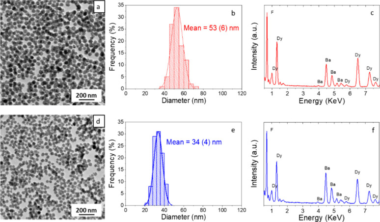
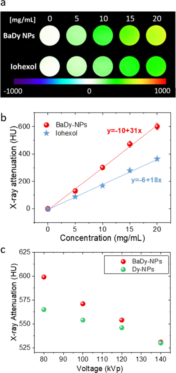
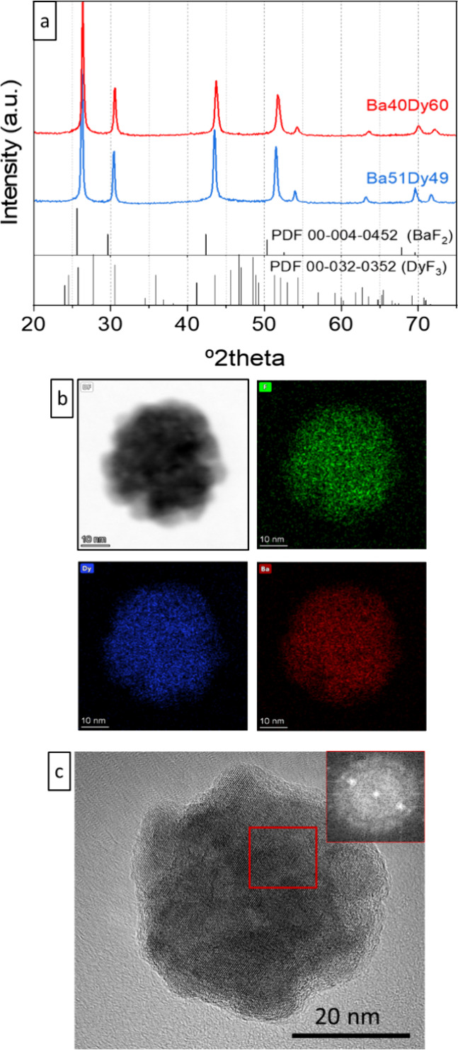
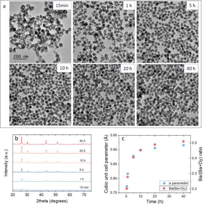
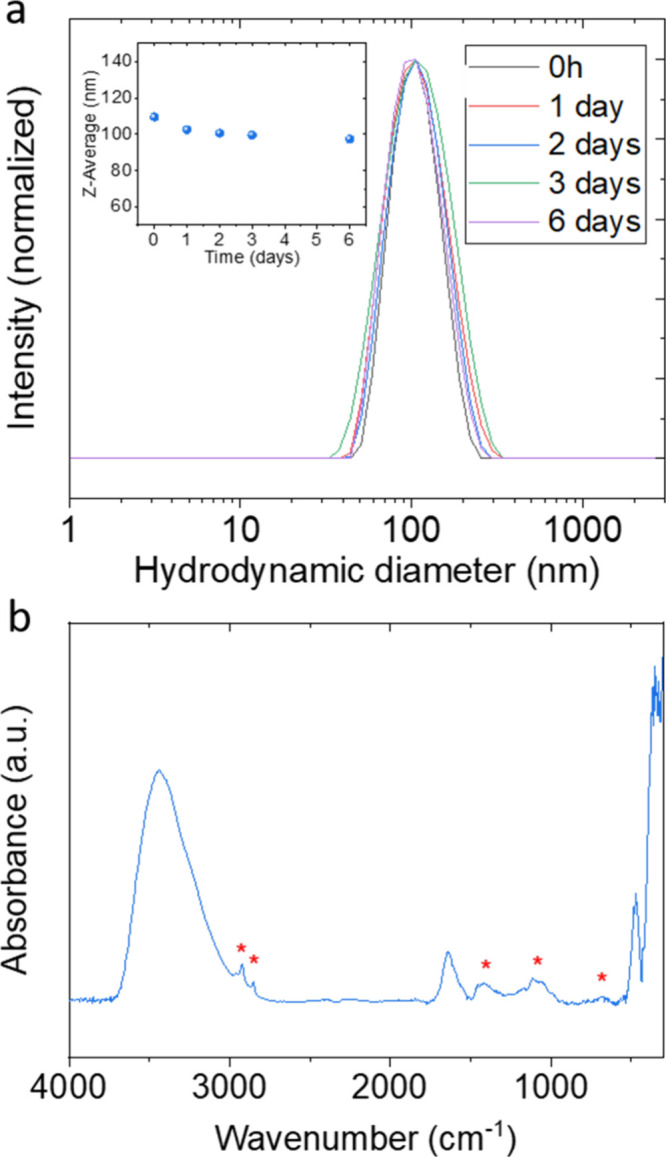
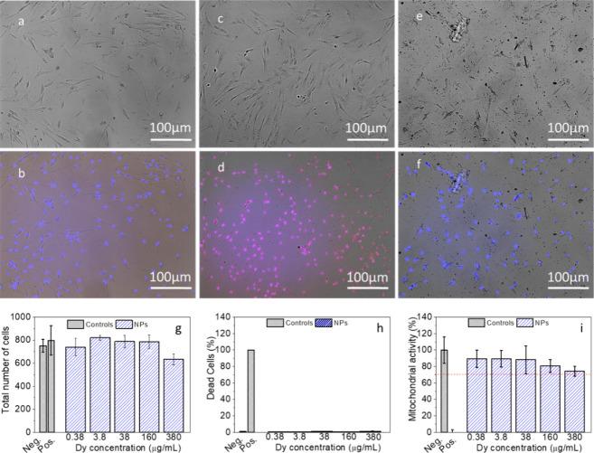
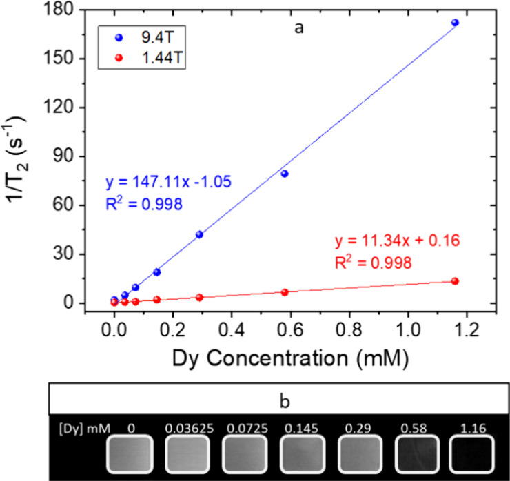
Bimodal medical imaging based on magnetic resonance imaging (MRI) and computed tomography (CT) is a well-known strategy to increase the diagnostic accuracy. The most recent advances in MRI and CT instrumentation are related to the use of ultra-high magnetic fields (UHF-MRI) and different working voltages (spectral CT), respectively. Such advances require the parallel development of bimodal contrast agents (CAs) that are efficient under new instrumental conditions. In this work, we have synthesized, through a precipitation reaction from a glycerol solution of the precursors, uniform barium dysprosium fluoride nanospheres with a cubic fluorite structure, whose size was found to depend on the Ba/(Ba + Dy) ratio of the starting solution. Moreover, irrespective of the starting Ba/(Ba + Dy) ratio, the experimental Ba/(Ba + Dy) values were always lower than those used in the starting solutions. This result was assigned to lower precipitation kinetics of barium fluoride compared to dysprosium fluoride, as inferred from the detailed analysis of the effect of reaction time on the chemical composition of the precipitates. A sample composed of 34 nm nanospheres with a BaDyF stoichiometry showed a transversal relaxivity () value of 147.11 mM·s at 9.4 T and gave a high negative contrast in the phantom image. Likewise, it produced high X-ray attenuation in a large range of working voltages (from 80 to 140 kVp), which can be attributed to the presence of different K-edge values and high Z elements (Ba and Dy) in the nanospheres. Finally, these nanospheres showed negligible cytotoxicity for different biocompatibility tests. Taken together, these results show that the reported nanoparticles are excellent candidates for UHF-MRI/spectral CT bimodal imaging CAs.
基于磁共振成像(MRI)和计算机断层扫描(CT)的双模态医学成像是提高诊断准确性的一种众所周知的策略。MRI和CT仪器的最新进展分别与超高磁场(UHF-MRI)的使用和不同的工作电压(光谱CT)有关。这些进展需要并行开发在新仪器条件下有效的双模态造影剂(CAs)。在这项工作中,我们通过从前体的甘油溶液中进行沉淀反应,合成了具有立方萤石结构的均匀氟化钡镝纳米球,发现其尺寸取决于起始溶液的Ba/(Ba + Dy) 比率。此外,无论起始Ba/(Ba + Dy) 比率如何,实验测得的Ba/(Ba + Dy) 值总是低于起始溶液中使用的值。从对反应时间对沉淀物化学成分影响的详细分析推断,该结果归因于氟化钡的沉淀动力学低于氟化镝。一个由化学计量比为BaDyF的34 nm纳米球组成的样品在9.4 T时的横向弛豫率()值为147.11 mM·s,并且在体模图像中给出了高负对比度。同样,它在大范围的工作电压(80至140 kVp)下产生了高X射线衰减,这可归因于纳米球中存在不同的K边值和高Z元素(Ba和Dy)。最后,这些纳米球在不同的生物相容性测试中显示出可忽略不计的细胞毒性。综上所述,这些结果表明所报道的纳米颗粒是UHF-MRI/光谱CT双模态成像造影剂的极佳候选物。