Sato Yukiko, Ueda-Arakawa Naoko, Takahashi Ayako, Miyake Masahiro, Mori Yuki, Miyara Yasunori, Hara Chikako, Kitajima Yoko, Maruko Ruka, Kawai Moeko, Takahashi Hajime, Koizumi Hideki, Maruyama-Inoue Maiko, Yanagi Yasuo, Iida Tomohiro, Takahashi Kanji, Sakamoto Taiji, Tsujikawa Akitaka
Department of Ophthalmology and Visual Sciences, Kyoto University Graduate School of Medicine, Kyoto, Japan.
Department of Ophthalmology, Graduate School of Medicine, University of the Ryukyus, Nishihara, Okinawa, Japan.
Ophthalmol Sci. 2024 Apr 10;4(5):100528. doi: 10.1016/j.xops.2024.100528. eCollection 2024 Sep-Oct.
To elucidate the clinical characteristics and progression rates of pachychoroid and conventional geographic atrophy (GA).
Retrospective, multicenter, observational study.
A total of 173 eyes from 173 patients (38 eyes with pachychoroid GA and 135 with conventional GA) from 6 university hospitals in Japan were included. All patients were Japanese, aged ≥50 years and with fundus autofluorescence images having analyzable image quality. A total of 101 eyes (22 with pachychoroid GA and 79 with conventional GA) were included in the follow-up group.
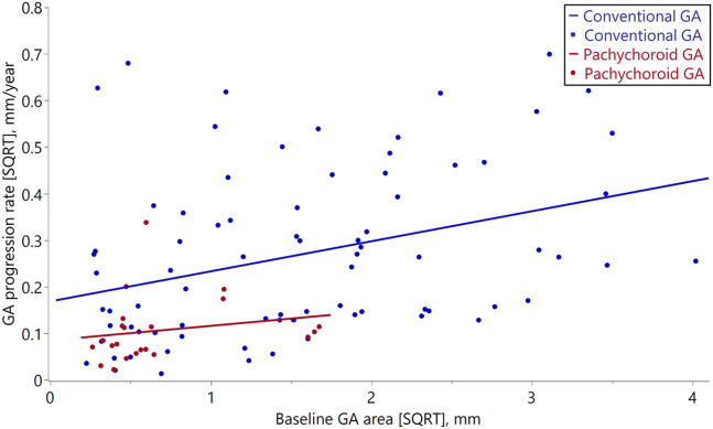
The studied eyes were classified as having pachychoroid or conventional GA; the former was diagnosed if the eye had features of pachychoroid and no drusen. The GA area was semiautomatically measured on fundus autofluorescence images, and the GA progression rate was calculated for the follow-up group. Multivariable linear regression analysis was used to determine whether the rate of GA progression was associated with GA subtype.
Clinical characteristics and progression rates of pachychoroid and conventional GA.
The pachychoroid GA group was significantly younger (70.3 vs. 78.7 years; < 0.001), more male-dominant (89.5 vs. 55.6%; < 0.001), and had better best-corrected visual acuity (0.15 vs. 0.40 in logarithm of the minimum angle of resolution; = 0.002), thicker choroid (312.4 vs. 161.6 μm; < 0.001), higher rate of unifocal GA type (94.7 vs. 49.6%; < 0.001), and smaller GA area (0.59 vs. 3.76 mm < 0.001) than the conventional GA group. In the follow-up group, the mean GA progression rate (square-root transformation) was significantly lower in the pachychoroid GA group than in the conventional GA group (0.11 vs. 0.27 mm/year; < 0.001).
Demographic and ocular characteristics differed between GA subtypes. The progression rate of pachychoroid GA, adjusted for age and baseline GA area, was significantly lower than that of conventional GA. Japanese patients with conventional GA showed characteristics and progression rates similar to those in White populations. Some characteristics of GA in Japanese population differ from those in Waucasian populations, which may be due to the inclusion of pachychoroid GA.
Proprietary or commercial disclosure may be found in the Footnotes and Disclosures at the end of this article.
阐明厚脉络膜型和传统地图样萎缩(GA)的临床特征及进展速度。
回顾性、多中心、观察性研究。
纳入了来自日本6所大学医院的173例患者的173只眼(38只眼为厚脉络膜型GA,135只眼为传统GA)。所有患者均为日本人,年龄≥50岁,且眼底自发荧光图像具有可分析的图像质量。共有101只眼(22只眼为厚脉络膜型GA,79只眼为传统GA)纳入随访组。
将研究的眼睛分为厚脉络膜型或传统GA;若眼睛具有厚脉络膜特征且无玻璃膜疣,则诊断为前者。在眼底自发荧光图像上半自动测量GA面积,并计算随访组的GA进展速度。采用多变量线性回归分析确定GA进展速度是否与GA亚型相关。
厚脉络膜型和传统GA的临床特征及进展速度。
厚脉络膜型GA组患者显著更年轻(70.3岁对78.7岁;P<0.001),男性占比更高(89.5%对55.6%;P<0.001),最佳矫正视力更好(最小分辨角对数视力分别为0.15对0.40;P=0.002),脉络膜更厚(312.4μm对161.6μm;P<0.001),单灶性GA类型的比例更高(94.7%对49.6%;P<0.001),GA面积更小(0.59mm²对3.76mm²;P<0.001)。在随访组中,厚脉络膜型GA组的平均GA进展速度(平方根转换)显著低于传统GA组(0.11mm/年对0.27mm/年;P<0.001)。
GA亚型之间的人口统计学和眼部特征存在差异。在调整年龄和基线GA面积后,厚脉络膜型GA的进展速度显著低于传统GA。日本传统GA患者的特征和进展速度与白种人群相似。日本人群中GA的一些特征与白种人群不同,这可能是由于纳入了厚脉络膜型GA。
在本文末尾的脚注和披露中可能会发现专有或商业披露信息。