Morano Alexander A, Ali Ilzat, Dvorin Jeffrey D
Biological and Biomedical Sciences, Harvard Medical School, Boston, Massachusetts, United States of America.
Division of Infectious Diseases, Boston Children's Hospital, Boston, Massachusetts, United States of America.
PLoS Pathog. 2024 Jun 3;20(6):e1012265. doi: 10.1371/journal.ppat.1012265. eCollection 2024 Jun.
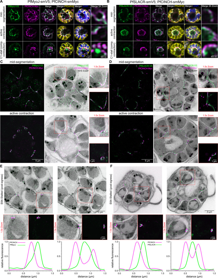
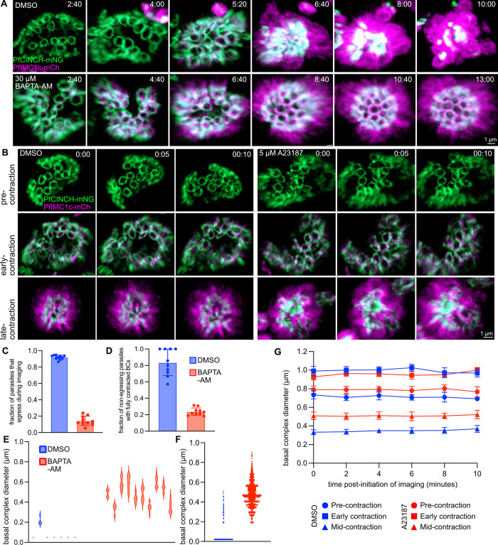
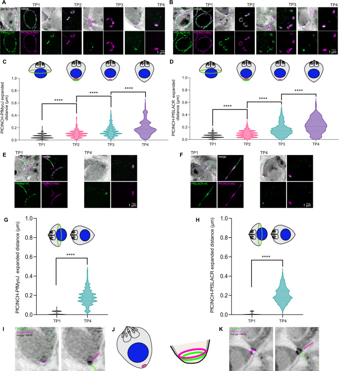
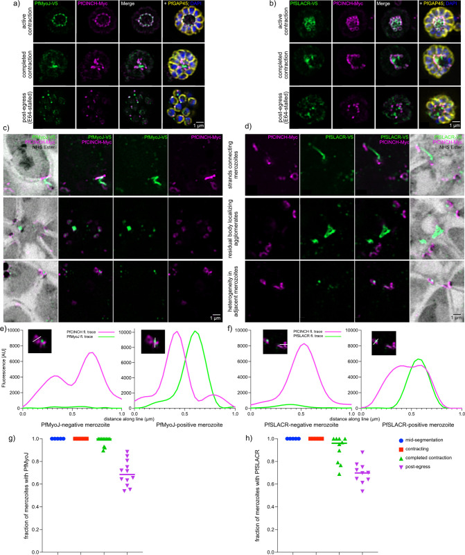
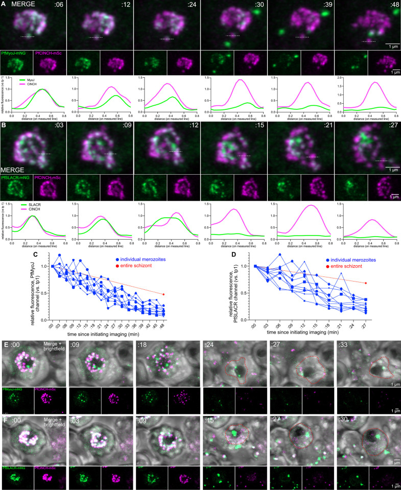
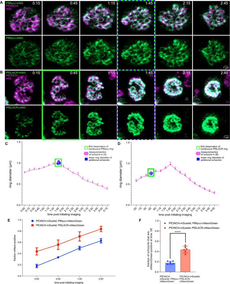
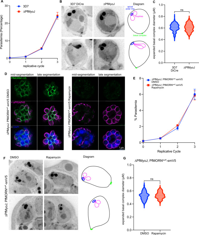
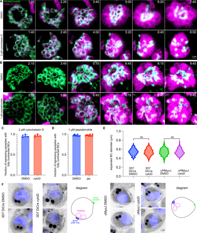
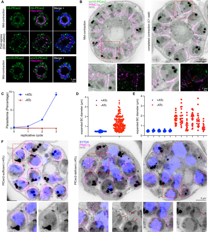
Asexual replication of Plasmodium falciparum occurs via schizogony, wherein 16-36 daughter cells are produced within the parasite during one semi-synchronized cytokinetic event. Schizogony requires a divergent contractile ring structure known as the basal complex. Our lab has previously identified PfMyoJ (PF3D7_1229800) and PfSLACR (PF3D7_0214700) as basal complex proteins recruited midway through segmentation. Using ultrastructure expansion microscopy, we localized both proteins to a novel basal complex subcompartment. While both colocalize with the basal complex protein PfCINCH upon recruitment, they form a separate, more basal subcompartment termed the posterior cup during contraction. We also show that PfSLACR is recruited to the basal complex prior to PfMyoJ, and that both proteins are removed unevenly as segmentation concludes. Using live-cell microscopy, we show that actin dynamics are dispensable for basal complex formation, expansion, and contraction. We then show that EF-hand containing P. falciparum Centrin 2 partially localizes to this posterior cup of the basal complex and that it is essential for growth and replication, with variable defects in basal complex contraction and synchrony. Finally, we demonstrate that free intracellular calcium is necessary but not sufficient for basal complex contraction in P. falciparum. Thus, we demonstrate dynamic spatial compartmentalization of the Plasmodium falciparum basal complex, identify an additional basal complex protein, and begin to elucidate the unique mechanism of contraction utilized by P. falciparum, opening the door for further exploration of Apicomplexan cellular division.
恶性疟原虫的无性繁殖通过裂体生殖进行,在此过程中,在一次半同步的细胞动力学事件中,寄生虫体内会产生16 - 36个 daughter 细胞。裂体生殖需要一种不同的收缩环结构,即基底复合体。我们实验室之前已鉴定出PfMyoJ(PF3D7_1229800)和PfSLACR(PF3D7_0214700)为在分裂中期被招募的基底复合体蛋白。利用超微结构扩展显微镜,我们将这两种蛋白定位到一个新的基底复合体亚区室。虽然在被招募时两者都与基底复合体蛋白PfCINCH共定位,但在收缩过程中它们形成一个单独的、更靠后的亚区室,称为后杯。我们还表明PfSLACR在PfMyoJ之前被招募到基底复合体,并且在分裂结束时这两种蛋白被不均匀地去除。利用活细胞显微镜,我们表明肌动蛋白动力学对于基底复合体的形成、扩展和收缩是可有可无的。然后我们表明含有EF手结构的恶性疟原虫中心蛋白2部分定位于基底复合体的这个后杯,并且它对于生长和复制是必不可少的,在基底复合体收缩和同步方面存在可变缺陷。最后,我们证明游离细胞内钙对于恶性疟原虫基底复合体的收缩是必要的,但不是充分的。因此,我们展示了恶性疟原虫基底复合体的动态空间分隔,鉴定出一种额外的基底复合体蛋白,并开始阐明恶性疟原虫利用的独特收缩机制,为进一步探索顶复门细胞分裂打开了大门。