Russell H. Morgan Department of Radiology and Radiological Science, Johns Hopkins University, Baltimore, MD, 21287, USA.
Institute for Cell Engineering, Johns Hopkins University, Baltimore, MD, 21218, USA.
Int J Nanomedicine. 2024 May 30;19:4995-5010. doi: 10.2147/IJN.S454128. eCollection 2024.
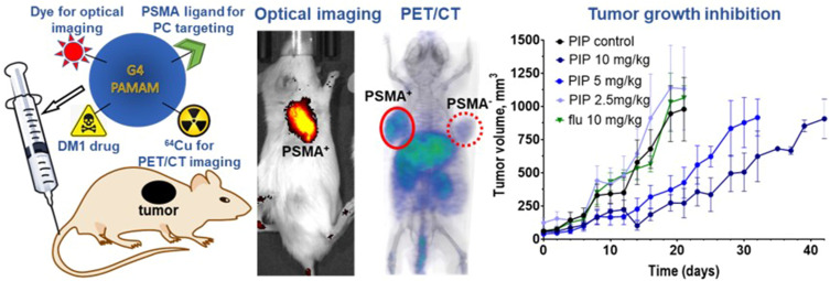
Prostate cancer (PC) is the second most common cancer and the fifth most frequent cause of cancer death among men. Prostate-specific membrane antigen (PSMA) expression is associated with aggressive PC, with expression in over 90% of patients with metastatic disease. Those characteristics have led to its use for PC diagnosis and therapies with radiopharmaceuticals, antibody-drug conjugates, and nanoparticles. Despite these advancements, none of the current therapeutics are curative and show some degree of toxicity. Here we present the synthesis and preclinical evaluation of a multimodal, PSMA-targeted dendrimer-drug conjugate (PT-DDC), synthesized using poly(amidoamine) (PAMAM) dendrimers. PT-DDC was designed to enable imaging of drug delivery, providing valuable insights to understand and enhance therapeutic response.
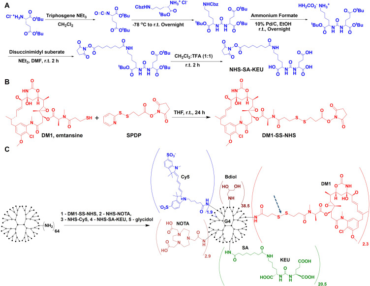
The PT-DDC was synthesized through consecutive conjugation of generation-4 PAMAM dendrimers with maytansinoid-1 (DM1) a highly potent antimitotic agent, Cy5 infrared dye for optical imaging, 2,2',2"-(1,4,7-triazacyclononane-1,4,7-triyl)triacetic acid (NOTA) chelator for radiolabeling with copper-64 and positron emission tomography tomography/computed tomography (PET/CT), lysine-urea-glutamate (KEU) PSMA-targeting moiety and the remaining terminal primary amines were capped with butane-1,2-diol. Non-targeted control dendrimer-drug conjugate (Ctrl-DDC) was formulated without conjugation of KEU. PT-DDC and Ctrl-DDC were characterized using high-performance liquid chromatography, matrix assisted laser desorption ionization mass spectrometry and dynamic light scattering. In vitro and in vivo evaluation of PT-DDC and Ctrl-DDC were carried out in isogenic human prostate cancer PSMA PC3 PIP and PSMA PC3 flu cell lines, and in mice bearing the corresponding xenografts.
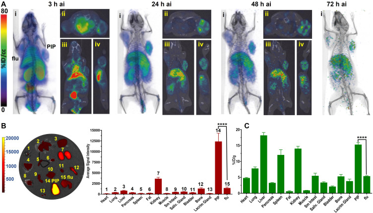
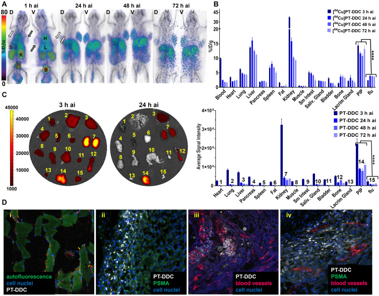
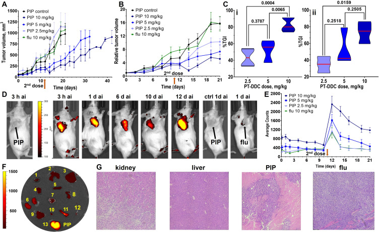
PT-DDC was stable in 1×PBS and human blood plasma and required glutathione for DM1 release. Optical, PET/CT and biodistribution studies confirmed the in vivo PSMA-specificity of PT-DDC. PT-DDC demonstrated dose-dependent accumulation and cytotoxicity in PSMA PC3 PIP cells, and also showed growth inhibition of the corresponding tumors. PT-DDC did not accumulate in PSMA PC3 flu tumors and did not inhibit their growth. Ctrl-DDC did not show PSMA specificity.
In this study, we synthesized a multimodal theranostic agent capable of delivering DM1 and a radionuclide to PSMA tumors. This approach holds promise for enhancing image-guided treatment of aggressive, metastatic subtypes of prostate cancer.
前列腺癌(PC)是男性中第二常见的癌症和第五大常见的癌症死亡原因。前列腺特异性膜抗原(PSMA)的表达与侵袭性 PC 相关,在超过 90%的转移性疾病患者中表达。这些特征使其可用于 PC 的放射性药物、抗体药物偶联物和纳米颗粒的诊断和治疗。尽管取得了这些进展,但目前的治疗方法都没有治愈效果,并且具有一定程度的毒性。在这里,我们提出了一种多模态、PSMA 靶向树状聚合物-药物偶联物(PT-DDC)的合成和临床前评估,该偶联物是使用聚(酰胺-胺)(PAMAM)树状聚合物合成的。PT-DDC 的设计旨在实现药物递送的成像,为理解和增强治疗反应提供有价值的见解。
PT-DDC 是通过连续接枝于四代 PAMAM 树状聚合物上合成的,其中包括美坦新-1(DM1)——一种高效的抗有丝分裂剂、Cy5 红外染料用于光学成像、2,2',2"-(1,4,7-三氮杂环壬烷-1,4,7-三基)三乙酸(NOTA)螯合剂用于标记铜-64 和正电子发射断层扫描/计算机断层扫描(PET/CT)、赖氨酸-脲-谷氨酸(KEU)PSMA 靶向部分和剩余的末端伯胺用丁烷-1,2-二醇封闭。未连接 KEU 合成了非靶向对照树状聚合物-药物偶联物(Ctrl-DDC)。PT-DDC 和 Ctrl-DDC 采用高效液相色谱法、基质辅助激光解吸电离质谱法和动态光散射进行表征。在同源人前列腺癌 PSMA PC3 PIP 和 PSMA PC3 flu 细胞系以及携带相应异种移植物的小鼠中进行了 PT-DDC 和 Ctrl-DDC 的体外和体内评价。
PT-DDC 在 1×PBS 和人血浆中稳定,需要谷胱甘肽才能释放 DM1。光学、PET/CT 和生物分布研究证实了 PT-DDC 的体内 PSMA 特异性。PT-DDC 在 PSMA PC3 PIP 细胞中表现出剂量依赖性的积累和细胞毒性,并且还抑制了相应肿瘤的生长。PT-DDC 不会在 PSMA PC3 flu 肿瘤中积累,也不会抑制其生长。Ctrl-DDC 没有表现出 PSMA 的特异性。
在这项研究中,我们合成了一种多模态治疗剂,能够向 PSMA 肿瘤递送 DM1 和放射性核素。这种方法有望增强对侵袭性、转移性前列腺癌亚型的图像引导治疗。